Outer part of the hand. The structure of the bone composition of the hand. Structure of the hand: metacarpophalangeal joints
The muscles of the hand are located mainly on the palmar surface of the hand and are divided into the lateral group (muscles of the thumb), medial group (muscles of the little finger) and the middle group. On the dorsum of the hand are the dorsal (back) interosseous muscles.
Lateral group
The short muscle that abducts the thumb (m. abductor pollicis brevis) (Fig. 120, 121) abducts the thumb, slightly opposing it, and takes part in flexion of the proximal phalanx. It is located directly under the skin on the lateral side of the eminence of the thumb. It begins on the scaphoid bone and ligament of the palmar surface of the wrist, and is attached to the lateral surface of the base of the proximal phalanx of the thumb.
| Rice. 120. Muscles of the hand (palm surface): 1 - pronator quadratus; |
|
 | Rice. 121. Muscles of the hand (palm surface): 1 - pronator quadratus; |
 | Rice. 122. Muscles of the hand (dorsal surface): |
 | Rice. 123. Muscles of the hand (dorsal surface): 1 - short extensor pollicis; |
The short flexor pollicis brevis (m. flexor pollicis brevis) (Fig. 120, 121) flexes the proximal phalanx of the thumb. This muscle is also located just under the skin and has two heads. The starting point of the superficial head is on the ligamentous apparatus of the palmar surface of the wrist, and the deep head is on the trapezius bone and the radiate ligament of the wrist. Both heads are attached to the sesamoid bones of the metacarpophalangeal joint of the thumb.
The muscle opposing the thumb to the hand (m. opponens pollicis) (Fig. 120, 121) opposes the thumb to the little finger. It is located under the abductor pollicis brevis muscle and is a thin triangular plate. The muscle starts from the ligamentous apparatus of the palmar surface of the wrist and the tubercle of the costoptrapezium, and is attached to the lateral edge of the first metacarpal bone.
The muscle that adducts the thumb (m. adductor pollicis) (Fig. 120, 123) adducts the thumb and takes part in the flexion of its proximal phalanx. It lies the deepest of all the muscles of the eminence of the thumb and has two heads. The starting point of the transverse head (caput transversum) is located on the palmar surface of the IV metacarpal bone, the oblique head (caput obliquum) is on the capitate bone and the radiate ligament of the wrist. The attachment point for both heads is located at the base of the proximal phalanx of the thumb and the medial sesamoid bone of the metacarpophalangeal joint.
Medial group
The short palmar muscle (m. palmaris brevis) stretches the palmar aponeurosis, forming folds and dimples in the skin in the area of the eminence of the little finger. This muscle, which is a thin plate with parallel fibers, is one of the few cutaneous muscles available in humans. It has a point of origin on the inner edge of the palmar aponeurosis and the ligamentous apparatus of the wrist. The place of its attachment is located directly in the skin of the medial edge of the hand at the eminence of the little finger.
The muscle that abducts the little finger (m. abductor digiti minimi) (Fig. 122, 123) abducts the little finger and takes part in the flexion of its proximal phalanx. It is located under the skin and is partially covered by the palmaris brevis muscle. The muscle originates from the pisiform bone of the wrist and attaches to the ulnar edge of the base of the proximal phalanx of the little finger.
The short flexor of the little finger (m. flexor digiri minimi) bends the proximal phalanx of the little finger and takes part in its adduction. It is a small, flattened muscle covered by skin and partly by the palmaris brevis muscle. Its point of origin is located on the hamate and ligaments of the wrist, and its attachment point is on the palmar surface of the base of the proximal phalanx of the little finger.
The muscle adducting the little finger (m. opponens digiti minimi) (Fig. 120) opposes the little finger to the thumb. The outer edge of the muscle is covered by the short flexor of the little finger. It begins on the hamate and ligamentous apparatus of the wrist, and is attached to the ulnar edge of the fifth metacarpal bone.
Middle group
Vermiform muscles (mm. lumbricales) (Fig. 120, 123) bend the proximal phalanges of the II–V fingers and straighten their middle and distal phalanges. There are four muscles in total, all of them have a spindle-shaped shape and are directed to the II–IV fingers. All four muscles begin from the radial edge of the corresponding tendon of the deep flexor digitorum, and are attached to the dorsal surface of the base of the proximal phalanges of the II–IV fingers.
The palmar interosseous muscles (mm. interossei palmares) (Fig. 120, 121) flex the proximal phalanges, extend the middle and distal phalanges of the little finger, index and ring fingers, simultaneously bringing them to the middle finger.
They are located in the interosseous spaces between the II–V metacarpal bones and represent three muscle bundles. The first interosseous muscle is located on the radial half of the palm, its origin point is the medial side of the II metacarpal bone, the second and third interosseous muscles are located on the ulnar half of the palm, their origin point is the lateral side of the IV and V metacarpal bones. The place of muscle attachment is the base of the proximal phalanges of the II–V fingers and the articular capsules of the metacarpophalangeal joints of the same fingers.
Dorsal interosseous muscles (mm. interossei dorsales) (Fig. 120, 121, 122, 123) flex the proximal phalanges, extend the distal and middle phalanges, and also abduct the little finger, index and ring fingers from the middle finger. They are the muscles of the dorsal surface of the hand. This group consists of four fusiform bipennate muscles, which are located in the interosseous spaces of the dorsum of the hand. Each muscle has two heads, which begin from the lateral surfaces of two adjacent metacarpal bones facing each other. The place of their attachment is the base of the proximal phalanges of the II–IV fingers. The first and second muscles are attached to the radial edge of the index and middle fingers, and the third and fourth are attached to the ulnar edge of the middle and ring fingers.
If we consider the hand as a whole, then, as in any other part of the human musculoskeletal system, three main structures can be distinguished: the bones of the hand; ligaments of the hand, which hold the bones and form joints; muscles of the hand.
Hand bones
The hand has three sections: wrist, metacarpus and fingers.
Carpal bones The eight small bones of the wrist have an irregular shape. They are located in two rows.
The proximal row consists of the following bones, if you go from the thumb to the fifth finger: scaphoid, lunate, triquetrum and pisiform.
The distal row also consists of four bones: polygonal, trapezoid, capitate and hamate, which with its hook faces the palmar side of the hand.
The proximal row of carpal bones forms an articular surface convex towards the radius. The distal row is connected to the proximal row using an irregularly shaped joint.
The bones of the wrist lie in different planes and form a groove (carpal groove) on the palmar surface and a bulge on the back. The groove of the wrist contains the tendons of the finger flexor muscles. Its inner edge is limited by the pisiform bone and the hook of the hamate bone, which are easily palpable; the outer edge is composed of two bones - the scaphoid and the polygonal.

Metacarpal bones
The metacarpus consists of five tubular metacarpal bones. The metacarpal bone of the first finger is shorter than the others, but is distinguished by its massiveness. The longest is the second metacarpal bone. The following bones towards the ulnar edge of the hand decrease in length. Each metacarpal bone has a base, a body and a head.
The bases of the metacarpal bones articulate with the bones of the wrist. The bases of the first and fifth metacarpal bones have saddle-shaped articular surfaces, and the rest have flat articular surfaces. The heads of the metacarpal bones have a hemispherical articular surface and articulate with the proximal phalanges of the fingers.
Finger bones
Each finger consists of three phalanges: proximal, middle and distal. The exception is the first finger, which has only two phalanges - proximal and distal. The proximal phalanges are the longest, the distal ones are the shortest. Each phalanx has a middle part - a body and two ends - proximal and distal. At the proximal end is the base of the phalanx, and at the distal end is the head of the phalanx. At each end of the phalanx there are articular surfaces for articulation with adjacent bones.
Sesamoid bones of the hand
In addition to these bones, the hand also has sesamoid bones, which are located in the thickness of the tendons between the metacarpal bone of the thumb and its proximal phalanx. There are also unstable sesamoid bones between the metacarpal bone and the proximal phalanx of the second and fifth fingers. Sesamoid bones are usually located on the palmar surface, but are occasionally found on the dorsal surface. The sesamoid bones also include the pisiform bone. All sesamoid bones, as well as all processes of bones, increase the leverage of the muscles that are attached to them.
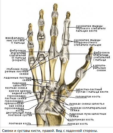
Ligamentous apparatus of the hand
Wrist joint
The formation of this joint involves the radius and bones of the proximal row of the wrist: scaphoid, lunate and triquetrum. The ulna does not reach the surface of the radiocarpal joint (it is “supplemented” by the articular disc). Thus, in the formation of the elbow joint, the ulna plays the largest role of the two forearm bones, and the radius plays the largest role in the formation of the radiocarpal joint.
In the radiocarpal joint, which has an elliptical shape, flexion and extension, adduction and abduction of the hand are possible. Pronation and supination of the hand occur together with the same movements of the bones of the forearm. A small passive rotational movement is also possible in the radiocarpal joint (10-12°), but this occurs due to the elasticity of the articular cartilage. The position of the gap of the radiocarpal joint is determined from the dorsal surface, where it is easily detected through the soft tissues; in addition, its position is determined from the radial and ulnar sides. On the radial side, in the area of the inferior radial fossa, you can palpate the gap between the lateral styloid process and the scaphoid bone. On the ulnar side, a depression is felt between the head of the ulna and the triquetral bone, corresponding to the ulnar portion of the cavity of the radiocarpal joint.
Movements in the radiocarpal joint are closely related to movements in the midcarpal joint, which is located between the proximal and distal rows of carpal bones. This joint has a complex, irregularly shaped surface. The total range of mobility when flexing the wrist reaches 85°, and when extending it is also approximately 85°. Adduction of the hand in these joints is possible by 40°, and abduction by 20°. In addition, circular movement (circumduction) is possible in the radiocarpal joint.
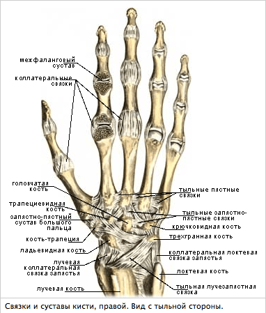
The radiocarpal and midcarpal joints are strengthened by numerous ligaments. The ligamentous apparatus of the hand is very complex. The ligaments are located on the palmar, dorsal, medial and lateral surfaces of the wrist, as well as between the individual bones of the wrist. The most important are the collateral ligaments of the wrist - the radial and ulnar. The first goes from the lateral styloid process to the scaphoid bone, the second - from the medial styloid process to the triquetral bone.
Between the bony elevations on the radial and ulnar sides of the palmar surface of the hand there is a ligament - the flexor retinaculum. It is not directly related to the joints of the hand, but is, in fact, a thickening of the fascia. Throwing over the carpal groove, it turns it into the carpal tunnel, where the flexor tendons of the fingers and the median nerve pass.
Carpometacarpal joints of the hand
They are connections of the distal row of carpal bones with the bases of the metacarpal bones. These joints, with the exception of the carpometacarpal joint of the thumb, are flat and inactive. The range of movements in them does not exceed 5-10°. Mobility in these joints, as well as between the bones of the wrist, is sharply limited by well-developed ligaments.
The ligaments located on the palmar surface of the hand make up a strong palmar ligamentous apparatus. It connects the carpal bones to each other, as well as to the metacarpal bones. On the hand you can distinguish ligaments that run arcuate, radial and transverse. The central bone of the ligamentous apparatus is the capitate, to which more ligaments are attached than to any other bone of the wrist. The dorsal ligaments of the hand are much less developed than the palmar ligaments. They connect the bones of the wrist to each other, making up thickening capsules covering the joints between these bones. In addition to the palmar and dorsal ligaments, the second row of carpal bones also has interosseous ligaments.
Due to the fact that the bones of the distal row of the wrist and the four (II-V) bones of the metacarpus are inactive relative to each other and are firmly connected into a single formation that makes up the central bone core of the hand, they are designated as the solid base of the hand.
The carpometacarpal joint of the thumb is formed by the polygonal bone and the base of the first metacarpal bone. The articular surfaces are saddle-shaped. The following movements are possible in the joint: adduction and abduction, opposition (opposition) and reverse movement (reposition), as well as circular movement (circumduction). Due to the opposition of the thumb to all other fingers, the scope of grasping movements of the hand increases significantly. The amount of mobility in the carpometacarpal joint of the thumb is 45-60° during abduction and adduction and 35-40° during opposition and reverse movement.
Metacarpophalangeal joints of the hand
Formed by the heads of the metacarpal bones and the bases of the proximal phalanges of the fingers. All these joints have a spherical shape and, accordingly, three mutually perpendicular axes of rotation, around which flexion and extension, adduction and abduction, as well as circular movement (circumduction) occur. Flexion and extension are possible at 90-100°, abduction and adduction - at 45-50°.
The metacarpophalangeal joints are strengthened by collateral ligaments located on the sides of them. On the palmar side, the capsules of these joints have additional ligaments called palmar ligaments. Their fibers are intertwined with the fibers of the deep transverse metacarpal ligament, which prevents the heads of the metacarpal bones from diverging to the sides.
Interphalangeal joints of the hand
They have a block-like shape, their axes of rotation run transversely. Flexion and extension are possible around these axes. Their volume in the proximal interphalangeal joints is 110-120°, while in the distal ones it is 80-90°. All interphalangeal joints are strengthened by well-defined collateral ligaments.
Fibrous and synovial sheaths of the tendons of the fingers
The flexor retinaculum and extensor retinaculum ligaments are of great importance for strengthening the position of the muscle tendons passing under them, especially when flexing and extending the hand: the tendons rest on the named ligaments from their inner surface, and the ligaments prevent the tendons from moving away from the bones and withstand significant pressure during strong muscle contractions .
The sliding of the tendons of the muscles passing from the forearm to the hand and the reduction of friction are facilitated by special tendon sheaths, which are fibrous or osteo-fibrous canals, inside of which there are synovial sheaths, which in some places extend beyond these canals. The largest number of synovial sheaths (6-7) is located under the extensor retinaculum. The formation of the canals involves the ulna and radius bones, which have grooves corresponding to the passage of the muscle tendons, and fibrous bridges that separate one canal from the other, which go from the extensor retinaculum to the bones.
The palmar synovial sheaths belong to the flexor tendons of the hand and fingers running in the carpal canal. The tendons of the superficial and deep flexor fingers lie in a common synovial sheath, which extends to the middle of the palm, reaching the distal phalanx of only the fifth finger, and the tendon of the flexor pollicis longus is located in a separate synovial sheath, which passes along with the tendon onto the finger. In the palm area, the tendons of the muscles going to the second, third and fourth fingers are deprived of synovial sheaths for some distance and receive them again on the fingers. Only the tendons leading to the fifth finger have a synovial sheath, which is a continuation of the common synovial sheath for the flexor tendons of the fingers.
Muscles of the hand

On the hand, the muscles are located only on the palmar side. Here they form three groups: the middle one (in the middle section of the palmar surface), the thumb muscle group and the small finger muscle group. The large number of short muscles on the hand is due to the fine differentiation of finger movements.
Middle hand muscle group
Consists of lumbrical muscles that originate from the tendons of the deep flexor digitorum and are attached to the base of the proximal phalanges of the second to fifth fingers; palmar and dorsal interosseous muscles, which are located in the interosseous spaces between the metacarpal bones and are attached to the base of the proximal phalanges of the second to fifth fingers. The function of the muscles of the middle group is that they are involved in flexing the proximal phalanges of these fingers. In addition, the palmar interosseous muscles bring the fingers of the hand towards the middle finger, and the dorsal interosseous muscles spread them apart.
Thumb muscle group
Forms the so-called eminence of the thumb on the hand. They begin on the nearby bones of the wrist and metacarpus. Among them are distinguished: the short muscle that abducts the pollicis, which is attached to its proximal phalanx; flexor pollicis brevis, which attaches to the external sesamoid bone located at the base of the proximal phalanx of the thumb; the opponus pollicis muscle, which goes to the first metacarpal bone; and the adductor pollicis muscle, which attaches to the internal sesamoid bone located at the base of the proximal phalanx of the thumb. The function of these muscles is indicated in the name of each muscle.
Small finger muscle group
Forms an elevation on the inside of the palm. This group includes: palmaris brevis; muscle that abducts the little finger; flexor little finger brevis and oppons little finger muscle. They arise from the nearby carpal bones and attach to the base of the proximal phalanx of the fifth finger and the fifth metacarpal bone. Their function is determined by the name of the muscles themselves. 

Materials used in the article: sportmedicine.ru
With the help of the joints of the upper and lower extremities, a person can perform a wide variety of movements and manipulations. The upper limb consists of the shoulder, forearm, shoulder girdle and hand. The joints of the hands allow you to explore the world around you. In order to know how this happens, you need to have an idea of the structure of the hand.
The arms are attached to the body by joints, muscles and the shoulder girdle. The shoulder girdle is considered the most active and powerful.
The ability to bend a limb creates special mobility and allows a person to perform many actions. It is with the help of the hand that a person makes familiar and necessary movements: for example, he takes a cup, writes with a pen, moves and controls his fingers.
Structure of the hand
The hand is connected to the forearm using the wrist and consists of the metacarpus, carpus and phalanx of the fingers. The hand contains 27 bones. The bones of the metacarpus and carpus join together to form the palm of the hand. The wrist consists of 8 spongy bones, which are arranged in two rows. Each row is represented by 4 short dice.
Top row:
- lunate bone;
- scaphoid;
- triangular bone;
- pisiform bone.
Bottom row:
- small trapezius;
- large trapezoid;
- capitate bone;
- hamate bone.
The metacarpus consists of five bones, the first of which is the flattest and shortest. All fingers on the hands consist of three phalanges, except the thumb: proximal phalanx, distal phalanx and middle phalanx. The thumb contains two phalanges: the main phalanx and the nail phalanx. Since the metacarpal bone is connected to the wrist through a joint, it allows a person to move the thumb from side to side from the other fingers.
All joints of the wrist, metacarpus and fingers are connected to each other using ligaments. A person can rotate the hand 180 degrees. The end of the radius and ulna, when connected to the wrist, form the radiocarpal joint, which can rotate in three axes.
Arm muscles
A person can make movements with the hand with the help of muscle contractions, most of which are long muscles: for example, the muscles of the forearm. Muscles are attached to bones by tendons. Tendons, in turn, are attached to ligaments and connective tissue. The hand contains the interosseous muscles, thenar muscles, hypothenar muscles and the lumbrical muscles.

The flexor muscles of the hand are innervated by the median and ulnar nerves, and the extensor muscles by the radial nerves. Blood enters the hand through the arteries: ulnar and radial. The branches of the arteries form a deep and superficial arch between themselves.
The skin of the hand contains a large number of sweat glands and nerve endings. The papillary layer contains Meissner's corpuscles, which are responsible for the sense of touch. Most of these bodies are found in the fingertips.
Muscle tendons are inserted into special canals, the walls of which are lined with synovial membrane. At the end there is a synovial sheath filled with a special fluid that acts as a lubricant and ensures the tendon glides during movement.
The upper limb is represented by the triceps and biceps brachii muscles, or in other words, the biceps and triceps. Such muscles are especially developed in athletes or people who often have to do physical work. The biceps is also connected by ligaments and tendons and performs flexion and extension of the arm. The triceps is located in the shoulder area on its back surface and is attached to the shoulder blade. The muscle tendon contains the synovial bursa.
Upper extremity injuries lead to dysfunction and disability.
Man is not only an upright walking creature, but also possesses intelligence and culture. In the process of evolution, the limbs of mammals have constantly changed, giving rise to many variations: from paws and legs to wings and flippers, but only humans have found such a wide use for their hands. Human hands, unlike the limbs of animals, do not serve to move the body in space, but are independent tools designed not only to facilitate the existence of the organism and its survival, but also to develop creative skills, create and transform the world around us. The hands of human hands differ even from the hands of primates, which are closest to us in terms of development.
A lot of time has passed since primitive man learned to make stone axes. Today, hard physical labor is left to the responsibility of machines, and human hands continue to evolve, practicing more subtle activities - from embroidery and dance to playing musical instruments and sign language.
The upper limbs begin to form in the sixth week of pregnancy at the same time as the brain, major organs and face. The rudiments of the hands and fingers appear earlier than the fingers and toes. After a person is born, his hands begin to train and master new movements much earlier than other parts of the body. Innate reflexes appear, which then turn into more meaningful behavior. Doctors point out that the development of motor skills in a newborn’s hands is as important as the development of speech.
Up to a month, the child has only a grasping reflex - thanks to the sense of touch, he squeezes what falls into his palm and does not let go. By three months, this reflex weakens, the child can straighten his fingers and grasp an object with his entire palm, without using the thumb and forefinger yet. By six months, children can grasp objects with all fingers and hold them in both hands. As children approach the age of one year, the ability to grasp something with their fingers develops. At this age, they love to twirl objects in their hands, examine them, and learn to pick them up and throw them. At one year old, a small person can already handle small objects, taking them with two fingers, the thumb and forefinger. By the age of two, children can hold a spoon, pen, or other object, using their shoulder and elbow rather than their hand when moving. At three years of age, the wrist develops more strongly, and objects are grasped due to movement in it.
By the age of five, the development of fine motor skills begins - children actively draw, put together puzzles, cut paper with scissors, and hold mugs. By the age of six, the hand is already well developed, and the thumb is very mobile for fine movements and strong grips. When manipulating, the whole arm is involved, but depending on the purpose, more movements occur in the shoulder and forearm (active games) or hand and fingers (drawing, board games). Over the course of seven years, a child masters a gigantic number of movements and positions, many of which become reflexive, and at the same time, neural connections are built up in the brain, so training hand motor skills is very important for the full development of intelligence. This was known back in the days of ancient civilizations, devoting a lot of time to such activities as calligraphy, embroidery and weaving, drawing and playing musical instruments. How skillfully a person could work with his hands was used to judge his intelligence and even spirituality.
The structure of the human hand
The anatomy of the human hand is quite complex. Since this is a very mobile part of the body, it has many bones and joints, ligaments and muscles, and human fingers, unlike animal limbs, have nails. The skin of the hands also differs from the skin of the body, having specific folds and special sensitivity. So let's start with a review of the bones. The bones of the hand are divided into three sections: the wrist, which includes eight bones; the metacarpus, consisting of five long bones, and the fingers, numbering a total of fourteen phalanges. The phalanges are tubular bones, unlike the rest of the bones that make up the hand. All the bones are quite small and located very close to each other.

Due to the fact that there are many bones and they are small, the hand has such plasticity and mobility, but because of this, in case of injury, the performance of the hand is noticeably reduced. The bones of the wrist form both low-moving joints (for example, in the wrist area) and joints. The wrist joint has the shape of a semicircle and provides flexion and extension of the hand. This is a very complex joint that gives the hand a wide range of motion. The bones of the wrist are connected to each other by ligaments and form a strong joint that can withstand heavy loads when supported by the hand. At the base of the thumb is a saddle joint that allows movement in two axes, making the thumb very functional.
The bones of the phalanges have ball-and-socket joints, which allows the fingers to bend in only one plane. Because of this, fingers are very susceptible to injury. Unlike the other fingers, which have three phalanges, the thumb is shorter and consists of only two bones. The hand has a wide network of innervation, responsible for touch, movement, the functioning of the sebaceous and sweat glands and thermoregulation. In general, hands are closely related to a person’s mental state. In moments of stress, hands often shake, go numb, everything falls out of them, and a person loses the ability to control them fully. Also, during emotional outbursts, the palms may become cold or sweaty.

The ability of the hands to touch (tactile, thermal, pain sensitivity and pressure sensation) is also variable and depends on the work being performed. If the work is delicate and requires sensitivity, then it increases; if the work is rough, then the ability to touch weakens and the skin becomes rougher. The presence of many nerve arches in the hand makes the skin of the hand not only an instrument of creation, but also of knowledge.
There are three main nerves in the human hand: median, ulnar and radial - they are located differently for each person. Each nerve along its path “gives off branches,” gradually becoming thinner. Some of these branches will collect information, and some will transmit stimuli from the central nervous system.
There are few muscles in the hands. They are located on the palm inside and outside. The thumb has much more muscles than the rest. It is interesting that the fingers themselves, which are the most mobile element of the hands, have no muscles. The flexors and extensors of the fingers are located in the palm and forearms. If you look at the back of the hand with the fingers raised as high as possible, you can see the extensor ligaments stretching from the fingers to the wrist.

The vessels feeding the hand form a deep arterial arch and a wide network of veins; from the back they are very clearly visible in some people, literally sticking out. The skin on the palm of the hand is also special. It is wear-resistant, devoid of hair and sebaceous glands, and has many receptors, especially on the fingertips. Only the skin in the lip area and the tip of the tongue perceive sensations more acutely. There are also many sweat glands on the skin of the palms and soles. To prevent the skin from slipping and shaking when working with your hands (like the skin of the scalp, for example), the hand’s own fascia is located underneath it - a layer of very dense connective tissue that firmly fuses with the skin and thereby limits it (the skin) mobility.
The surface of the palm is divided by folds formed from repeated bending in typical places. According to some ideas, these lines on the hand can predict fate or identify diseases. Usually everyone has three main folds, which are called the lines of the mind, heart and life. But sometimes it happens that there is a clearly defined transverse fold in the palm, it is also called the siminian line (monkey fold), often it indicates abnormal development: Down's disease, congenital stuttering, heart defect. Palmists see in this line special character traits that allow its owner not to separate feelings and reason.

The human hand can generally take three main positions: “scoop” (when the palm and fingers are straightened), “hook” (when the palm is straight and the fingers are bent) and “grip” (when the palm and fingers are bent and the thumb is directed across the palm). In this case, the fingers themselves can be spaced apart or located nearby.
The functionality of the human hand cannot be overestimated, especially when it comes to activities that require many precise small movements. People use their hands not only for work, but also for communication. Sign language is both everyday non-verbal communication and an auxiliary means in explaining or clarifying what is being said, as well as an independent language used by the deaf and dumb.
Particular attention should be paid to human nails. Unlike claws, the nail plate is flat, fused with the finger and is not capable of extension, like the claws of most animals. Nails begin to form already at the end of the first trimester; they help the fingertips to remain hard and not change shape when touched or grabbed, help with itching, and protect the finger from damage. Nails grow throughout your life, constantly, and can be as long as you want, while the nail plate curls into a spiral as it dries.
The nail itself consists of three parts: the root, the body and the free edge. Moreover, the root is formed by living cells, and everything else is dead and keratinized. The root, or matrix, is located inside, part of it can be seen in the form of a light semicircle at the base of the nail. The matrix is very sensitive and is easily damaged, which can be expressed in the form of spots on the nail or its curvature. Over time it usually recovers.
The thickness of the nail is determined by genes, each person has their own. The nail owes its hardness to the protein keratin, which is somewhat different from the keratin of hair and skin, as it contains more sulfur. In addition to sulfur, nails contain calcium, zinc, phosphorus, selenium and chromium. The nail has a porous structure, so when it is in water it becomes soft and thickens, absorbing moisture. On top of the nail, at its base, there is a special fold of skin - the cuticle; it protects the nail crevice from dirt and infections. Its edge is formed by dead cells, which are easily damaged and can be painlessly removed. Often, due to severe damage to the cuticle, burrs appear, located on living tissue, which leads to inflammation.

Slow nail growth can occur during breastfeeding, diet, stress, metabolic disorders, circulatory problems or the use of low-quality cosmetics. Sometimes grooves or ribbing appear on the nail; this is most often a consequence of injuries to the nail root, iron deficiency anemia, or the presence of inflammatory processes in the body that worsen metabolism. With a lack of zinc, even transverse furrows may appear. Nails can also peel for many reasons, but most often this occurs due to chemical or mechanical damage (household chemicals or the habit of biting nails), allergies, fungal infections, lack of nutrients and vitamins.
In general, if the quality of the diet is low, nails are an excellent indicator. When there is a lack of minerals, the body first sacrifices what is least important and insensitive - tooth enamel, hair and nails. There are times when nails may be completely absent. This can happen due to genetic disorders, surgical removal, but in case of injury or severe infections, the nails can recover, but this does not happen quickly.
Some interesting facts about nails:
- During pregnancy and during menstruation, a woman's nails grow faster.
- Men's nails grow more slowly.
- Nails grow faster in summer and during sleep.
- The rate of nail growth depends on the length of the fingers - on the thumb and little finger, nails grow more slowly, as in general in people with shorter fingers. Toenails are thicker and grow more slowly.
- Nails grow faster on the right hand in right-handed people and on the left in left-handed people.
- Nails grow faster after viral infections, this is due to the process of cleansing and cell renewal.
- In Ancient Egypt, the aristocracy was distinguished by the dark color of its nails, which were painted with a special natural composition.
- In the 17th century in France, it was customary not to knock on the door, but to cross the long nail on the little finger.
- In China, the length of nails spoke of wealth and nobility; a person with long nails could not engage in menial labor. Long nails were also a sign of wisdom. They were often covered with gold. Short nails could be extended using rice paper.
- In Ancient Rus', nails and hair were considered guardians of strength and power. They shorn them on certain days and burned them. The sick person's nails were cut so that the disease would go away with them.
- In the Middle Ages in Europe, women with unnatural nail colors were accused of witchcraft.

Fingerprints and palm prints are another interesting topic to discuss. In most animals, the lines on the fingers are arranged randomly - in chimpanzees they are straight, and in humans they form intricate patterns reminiscent of labyrinths. Papillary lines appear at 18 weeks of pregnancy. There are only three genetic disorders where lines are not formed and the palms and fingers remain smooth: Naegeli-Francheschetti-Jadasson syndrome, dermatopathy and adermatoglyphia. In the first two cases, the fingerprints are indistinguishable due to skin pigmentation disorders; in the latter case, the fingertips simply remain smooth. Everyone knows that the pattern on the skin of the hands is strictly individual, unchangeable and practically indestructible; it is used in identifying a person, identifying pathologies, etc. So why in reality are there papillary lines on the hands?
As scientists have found, this partly contributes to increased friction when grasping and manipulating objects, partly the roughness of the upper layer of the epidermis prevents the appearance of large blisters during burns and injuries, and most importantly, the lines help to better feel material objects. When touched, some of the lines will always be parallel to the irregularities of the object, which enhances the sense of touch. Sweat glands are found in abundance at the tops of the lines, so hands are never perfectly dry and clean; handprints almost always remain. The prints of the left and right hands are completely different, so when fingerprints are taken, they are taken from both hands.
- Koala fingerprints are very similar to human ones, even experts can confuse them.
- On the gecko's feet there are special grooves formed by even smaller bristles, which themselves consist of microscopic hairs. Thanks to this, geckos can stick and stay on any smooth surfaces. This happens due to electrostatic interactions at the micro level.
- A person is able to regenerate the lost end of a finger if the injury does not affect the second phalanx. There are cases when the third phalanx with damaged tissues was surgically removed, and it grew back. The nail was restored, as were the papillary patterns. The condition is that the wound remains unsutured.
- The human hand has unique proportions: the first phalanx of the fingers is equal to the sum of the lengths of the second and third, the sizes of the phalanges are correlated according to the rule of the golden ratio.
- The skin on the ring finger is thinner, so blood is taken from it. But he is the most sensitive - he can feel irregularities of two tenths of a millimeter.
- The thumb indicates mental health; in people with Down syndrome it is underdeveloped or crooked.
- In Rus', the index finger was called a finger. In ancient Rome, the middle finger was called dirty because it was used to perform hygienic procedures, and the ring finger was called clean or medicinal because pharmacists used it to mix drugs. The middle finger in Europe was called the big one.

Hand injuries and diseases
Injuries to the hands occur frequently; in most cases, the wrist and wrist joint are affected, since they are not protected by muscles. Dislocations and sprains are common, especially among athletes. Finger fractures occur less frequently.
A fracture of the bones of the hand, if there are no obvious curvatures, is most easily identified using an x-ray. The damaged area is immobilized and cooled; if necessary, a splint is applied. When a hand is bruised, the hand swells greatly; the injury site is first cooled using decongestants and then warmed up to speed up recovery.
If you fall on your hand, dislocations occur, and the vessels and nerves in the wrist joint are compressed, which leads to pain, numbness and loss of mobility. Resetting a dislocated wrist is difficult and it is best not to try it yourself unless you have experience. In addition to the wrist, you can get a dislocation of the metacarpus when you fall on a clenched fist or with a very strong blow. In this case, the fingers lose mobility, and the hand swells on the back side. If you fall on a straightened hand, it is possible to dislocate your fingers at the phalangometacarpal joint; in this case, the bones are adjusted by a doctor under anesthesia.
In addition to bones, in different cases, ligaments, of which there are a lot in the hand, can also be damaged. If the ligaments are damaged, not only limited mobility may appear, but also mobility may occur where it should not exist, for example, a finger may begin to hyperextend or bend to the side. A ligament rupture is no less painful than a bone fracture; often a ligament injury entails dislocation of the nearest joint. If the ligaments in the palm that provide compression of the fingers are damaged, then a round object is placed in the hand and the victim is taken to the hospital, since in this case the ligaments will have to be sewn together by a surgeon.
In relation to the hand, a person can experience various diseases and pathologies. The most common are:
- Tendinitis, or inflammation of the ligaments. It is often an occupational disease for people whose fingers are in motion for a long time due to work: pianists, typists, seamstresses, typewriters, etc. It cannot be completely cured, so those who are sick are given therapy and are advised to refrain from dynamic loads on their fingers.
- Carpal tunnel syndrome is also the cause of monotonous manual work, sometimes hormonal disorders and rheumatoid arthritis. This compresses the median nerve, leading to pain, swelling and numbness. A person has to stretch a limb to restore its functionality. This happens more often in the morning. Treatment is carried out using a bandage and anti-inflammatory drugs.
- Osteoarthritis is a well-known disease that damages the cartilage of the joint; less commonly, the causes are improperly healed intra-articular fractures of the fingers and rheumatoid arthritis. Since the joints suffer, the person’s finger mobility is impaired and he cannot perform minor manipulations. Painful sensations accompany movements and go away when the hands remain at rest. Osteoarthritis is treated with physiotherapy, massage, gymnastics and anti-inflammatory drugs.
- Gouty arthritis is a consequence of salt deposition that occurs due to the consumption of large amounts of animal protein. Accompanied by acute pain in the joint with redness and fever. The patient is prescribed a diet and anti-inflammatory drugs.
- Rheumatoid arthritis affects small joints. It is dangerous because it can lead to deformation and complete immobilization. The disease is accompanied by pain, swelling, and immobility. The disease is treated with physiotherapy, gymnastics, hormonal drugs and anti-inflammatory drugs.
- Writing cramp occurs only when trying to write something. Hands become weak, tremble, fingers cramp. The cause may be either stress or diseases of the cervical spine (compression of the nerves). Treatment involves gymnastics, medicinal baths and visits to a psychotherapist.
- Snapping finger syndrome is common in people engaged in manual labor that involves the use of force. Excessive tension causes swelling of the joints, which leads to decreased mobility and pain. The fingers can be squeezed, but it is difficult to straighten, and when this happens, a distinct click is heard.
- Raynaud's syndrome involves loss of blood circulation in the extremities; not only the arms, but also the feet can be affected. The cause may be hypothermia, smoking, chemical poisoning, stress, prolonged vibration or hereditary vascular diseases. Symptoms occur sporadically. The fingers begin to turn white and numb at the ends, and after the attack ends, a tingling sensation is felt. The disease progresses over time, the main treatment is removal of the provoking factor. Vasodilators are prescribed; in later stages, surgery may be required.
Despite all of the above, the hands can and should be strengthened. Gymnastics, yoga, and any moderate physical activity will not only make bones and ligaments strong, but will also develop joint mobility and help strengthen the small muscles of the hands. There are special hand exercises aimed at improving motor skills, as well as many activities that help strengthen fingers, such as knitting, modeling or macrame.
On the palm of a person, as well as on the feet and on the ears, there are many points that help regulate the activity of the entire body. This knowledge comes from Tibetan medicine, where they believe that each finger and each area on the hand corresponds to a specific organ.

So the thumb is responsible for the lungs, bronchi, liver and heart, the index finger is for all digestive organs, the middle finger is for the circulatory system, the ring finger is for the nervous system, and the little finger is for the kidneys and small intestine. By massaging, pressing or applying acupuncture to these places, you can activate certain processes in the body.

For example, by acting on point number 1, you can calm your breathing, relieve a cough, and stimulate the thyroid gland.
Point 2 will eliminate anxiety, relieve stress, and help cope with headaches and pain in the stomach and pancreas.
Point 3 will help resist diseases of the excretory system, relieve back and muscle pain, relieve toothache, and eliminate heartburn. In addition, the effect on this point has a calming effect.
Point 4 improves blood circulation, helps reduce discomfort during menstruation, ease migraines, and has a beneficial effect on vision. It will also help with liver and gallbladder disorders. Acts as a tonic, invigorating agent.
Point 5 has a good effect on the condition of the skin, lungs and colon.
Point 6, located on the little finger, will help overcome uncertainty, doubt and anxiety; it has a beneficial effect on the functioning of the heart and small intestines. In addition, you can influence the point for sore throat and bone diseases.
Point 7 is associated with the pancreas and is beneficial for diabetics as it can help regulate sugar levels.
Point 8 generally affects the digestive system and is useful for problems with indigestion, bloating, and constipation. The dot in the center of the palm, next to the eighth, is a general indicator of health. If you press harder on it, the obvious pain will become a sign of an internal disease. Perhaps there is some hidden inflammatory process or metabolic disorder.
Point 9 is associated with the heart and endocrine system. Can help with hormonal disorders and abnormal metabolism.
There is also a control point between the tubercles of the middle and ring fingers, pain in which indicates problems of the genitourinary area.
Work with sensitive points should be carried out by specialists, since without the necessary knowledge and experience you can get the opposite effect and encounter problems that did not exist before.
General diagnosis of health status based on the appearance of the hands is very common in Eastern medicine. For example, red or yellow hands indicate liver problems. Red fingertips indicate digestive problems; if there is redness at the base of the thumb, then there are problems in the reproductive sphere. If brown spots appear on the back of the hand, then there is a problem with the gallbladder. Age-related pigmentation in people primarily appears on the hands, face and décolleté. If a marble pattern appears on the palms, then attention should be paid to the autonomic nervous system. Constantly wet palms or a crawling sensation indicate hormonal disorders and hyperfunction of the thyroid gland. Dry palms indicate insufficient thyroid function. Peeling of the skin on the back of the hand indicates a lack of vitamins A and D; if the skin becomes rough, then check the gallbladder. Also, problems with the gallbladder can manifest as roughening of the skin on the index finger.
The temperature of the hands can also tell a lot about the health of their owner. Ice brushes indicate poor blood circulation. Sometimes the little fingers can even go numb. If your thumb is numb, pay attention to your respiratory system. Hot palms will tell you that the liver is overloaded with toxins that are forced to exit through the sweat glands on the hands and feet. People often experience burning feet syndrome. If you have an itch on the side of your index finger on your right hand, then you need to take a closer look at the large intestine.

- Webbing between the fingers on the hands occurs four times more often in white people than in other races.
- On clean palms, germs die within 10 minutes, since sweat contains an antiseptic. Dirt on the hands clogs the sweat glands, which causes bacteria to multiply well on unwashed palms.
- The fourth part of the motor cortex of the brain is responsible only for hand movements.
- A paper cut is more painful, since it turns out to be superficial, thin and even (and even wounds are more difficult to heal), which causes almost no blood to be released. Because of this, damaged nerve endings are exposed to air and come into contact with oxygen, producing painful sensations.
- The movement of one thumb involves nine separate muscles and three nerves.
In addition to determining the state of health, one can also judge the human psyche and his physiological archetype by looking at the hands. Here is one interpretation:
1. Square palm and short fingers. Type of Earth.
Such people are persistent, responsible, love to work physically, are not afraid of monotonous or hard work, are in good health, not emotional, and feel better in nature than in the city. They prefer stability, tradition and predictability; they do not like innovation, haste and do not rely on chance. They do not tolerate mental stress, stress and a sedentary lifestyle well. In general, they have strong immunity, but can sometimes suffer from headaches.
2.Square palm and long fingers. Air type.
These people are outwardly extroverted - they communicate a lot, their interests are multifaceted - but, nevertheless, they need moments of solitude. These are active intellectuals who love to plan everything and do their work well in a short time. They can work physically if this work is creative. However, they tend to get excited about many things at the same time, are talkative, chaotic, and superficial. They do not tolerate monotony and monotony well. They need to learn concentration and perseverance. In terms of health, they have a fast metabolism, they are almost always in a state of mild permanent stress-shake-up, this is natural for them, the main thing is that the stress does not turn into chronic protracted stress. They do not tolerate cold well and are susceptible to colds and respiratory diseases, as well as limb injuries. You should learn not to rush and do everything more measuredly, without jerks. Great success is achieved in athletics.
3. Long palm and short fingers. Fire type.
Inspirers and pioneers, leaders and speakers. Generate new ideas. They have strong immunity. Stress-resistant. Always in communication and in the spotlight. They can work physically, but they don’t like it - monotony is like a punishment for them. Subject to emotional outbursts, after which exhaustion may occur. It is important to learn to calm down, control emotions and relax. It is also important to avoid heavy foods, various stimulants (energy drinks, black coffee, etc.) and alternate physical activity with relaxation (for example, the gym and massage). Susceptible to cardiovascular diseases (hypertension, arrhythmia) and gastrointestinal diseases, you should avoid overheating the body (in a sauna or when staying in the sun in the heat).
4. Oblong palm and long fingers. Type of Water.
Creative personalities are very vulnerable, emotional and suspicious. They live by feelings and intuition. They can do light physical labor, but do not tolerate heavy physical labor. They cannot stand stressful situations, haste or competition. They feel good in a protected environment. It is important to learn to control emotions and avoid apathy and despondency. Cultivate self-confidence and will. They lose and gain weight easily, their metabolism is quite fast. They may suffer from endocrine disorders, as well as mental illnesses and sleep disorders. The state of health in general depends on the emotional background; during times of stress, the immune system suffers greatly. Susceptible to viral infections. It is useful to spend more time in nature, jogging, swimming, cycling, dancing, yoga and any aerobic exercise.
In conclusion, we can summarize that the hand is not only a delicate instrument for manipulation, but also a sensory instrument, an indicator of health, and a reflection of human character. With our hands we create objects of the material world, we cognize them, communicate, express ourselves and learn hidden character traits and even destiny. According to Eastern wisdom, there are additional chakras on the palms; with the help of hands you can heal and control the flow of external and internal energies. According to ancient descriptions, the Buddha's palms bore imprints of the Wheel of Dharma, a symbol of the teachings of Enlightenment, and the Hamsa - an open palm facing downwards - is a powerful protective amulet in many religions. Turned up, it meant boundless generosity and was associated with the wise Buddha. A palm with an image of an eye or a cross in it is a symbol of divine or mystical powers. The five fingers in many teachings symbolize the five senses, and in Islam - the five main saints. Thus, for a person, his hands are not only a part of his body, but also a way to gain a relationship with higher powers and comprehend the truth.

Our body has a complex structure. This includes bones, muscles, nerves, tendons, blood, and so on. Today we will get acquainted with the structural features of the human hand.
Hand bones
Human skeleton– a very complex system in which everything is interconnected. As we know, the human body consists of bones. In scientific terms, bone is, first of all, an independent structural organ.
The bones contain:
- Organic(about twenty-eight percent) these include proteins, fats, monosaccharides
- Inorganic substances: water, about fifty percent, salt, about twenty-two percent. There are also compounds of calcium, magnesium, cobalt, and fluorine.
Organic substances in the composition give elasticity to bones. With age, their number decreases and bones become fragile. Therefore, you should maintain your bones until old age by taking vitamins containing magnesium and calcium for bones.
The bones themselves are divided into three sections:
Wrist bones. Eight pieces, four on each level.
- First: Triangular, lunate, scaphoid, pisiform.
- Second: The bone is trapezoid, trapezoid, hamate and capitate.
Metacarpal bones: Five in number. Moreover, they are short and tubular.
Divided into departments:
- Base;
- Body;
- Head.
The phalanges are tubular in appearance. Long bones contain red bone marrow
Divided into parts:
- Main;
- Average;
- Elbow.
The unique composition of the cream is a source of important building elements for joints. Effective in the fight against many joint diseases.
Ideal for both prevention and treatment at home. Has antiseptic properties. Relieves swelling and pain, prevents salt deposition.
Joints of the hand
Joints- These are the connections of the bones of the human body. Let's get acquainted with the components of joints.
- Joint cavity;
- Surfaces for attachment to bones;
- Capsule.
There are types of joints in the hand:
- Radiocarpal;
- Carpometacarpal;
- Metacarpophalangeal.
First joint looks like a connection of the radius with the triquetrum, scaphoid and lunate. Thanks to the functioning of our joints, the movement of the hand is carried out. We will talk about it in detail below.
 At the carpophalangeal joint movement is limited due to the well-developed ligamentous apparatus. A significant number of ligaments attach to the capitate bone. The palmar ligaments have a higher degree of development than the dorsal ligaments.
Provides a ten degree stroke. Together they create the palmar-ligamentous apparatus. This joint at the thumb is separate from the other joints. Because of this, he is different from others.
During bending, the thumb can move towards the palm, which other fingers cannot do. Our thumb is very flexible. It can rotate sixty degrees on its axis.
The metacarpophalangeal joints are formed by phalanges. The heads themselves are spherical in shape. The fingers are driven and retracted. Consist of two ligaments. Movements are performed around two axes at ninety degrees. Among other things, the fingers can make circular movements.
The importance of joints in the body is to ensure the movement of body parts and hold bones together. Athletes often injure their joints, so consult a doctor if necessary.
Can't cope with joint pain?
Joint pain can appear at any age; it gives a person unpleasant sensations and often severe discomfort.
Do not allow joint diseases to develop, take care of them today!
It has the following properties:
- Relieves pain syndrome
- Promotes regeneration of cartilage tissue
- Effectively relieves muscle hypertonicity
- Fights swelling and eliminates inflammation
Ligaments of the hand
Our ligaments protect and support our bones. They are “helpers” of the joints. Joints provide bone movement and protect them from mechanical damage. Ligaments can become stretched as a result of injury from a fall. A large number of ligaments creates a fibrous layer.
On the hand, the ligamentous apparatus is represented by several types of ligaments:
- Rear;
- Palmar;
- Interarticular;
- Collateral.
Sprains of the ligamentous apparatus often occur.
Doctors characterize them by degree:
- Lightweight– minor tear, no difficulty moving the hand. The pain is mild
- Average– a small tear of the ligaments. Swelling on the wrist, movements are painful.
- Heavy- ligament rupture. The pain is acute, inflammation often occurs. A characteristic sign will be a hematoma.
If a person has injured a ligamentous apparatus, then he needs first aid. It is important to know that the faster help is provided, the faster the ligament recovery will be.
First aid for sprains:
- Cold compress for two hours.
- Fix the joint (splint, bandage).
- Apply anesthetic ointment.
There are several ways to treat sprains, for example:
- Ointments;
- Bandages;
- Physiotherapy;
- Folk remedies (porridge compress, vodka compress).
Muscles of the hand
The human body consists of muscles. The brush is no exception. A muscle is an active part of the human musculoskeletal system.
The muscles of the hand are divided into groups:
- Lateral;
- Medial;
- Average.
Each group will list the muscles, their location and the role they perform.
Lateral group
- Abductor pollicis brevis- branches off the thumb; also with the help of this muscle, the finger can perform a slightly opposing movement. Located on the side of the thumb.
- Flexor pollicis brevis– bends the phalanx of the thumb. Attached with its heads to the metacarpophalangeal joint.
- Oppons pollicis muscle– opposes the thumb.
- Adductor pollicis muscle- leads the finger. Located under the short muscles.
Medial group
- The muscle that abducts the little finger is the abductor of the little finger. Located under the skin.
- Flexor pollicis brevis – flexes the little finger. Located under the skin.
- The muscle that adducts the little finger – adducts the little finger. Located under the short flexor.
Middle group
The middle group includes muscles that are worm-shaped. They start from the flexor muscles. They have one function - thanks to us we can bend the phalanges.
 Stories from our readers!
“I ordered the cream for myself for prevention and for my mother for the treatment of joints. Both were completely delighted! The composition of the cream is impressive, everyone has long known how useful and, most importantly, effective beekeeping products are.
After 10 days of use, my mother’s constant pain and stiffness in her fingers subsided. My knees stopped bothering me. Now this cream is always in our house. We recommend."
Characteristics of movements in the wrist joint
Let us dwell in more detail on the features of the elbow joint. The elbow joint, like all joints, is strengthened by ligaments.
The joint has its own characteristics:
- Ellipse shape. Thanks to this shape, it is capable of movement in both axes
- It has a complex articulation.
- Contains additional cartilage, thanks to which the conformity of the shape to the articular surfaces is ensured.
Scientists argued that the movement of the brush is circular. This is true, but it is ensured by the movement of not one joint, as they thought, but several: the radiocarpal, carpometacarpal, midcarpal, intercarpal joints.
Normally, movements in the wrist joint range from seventy-five to eighty degrees. Lateral movements up to twenty degrees
Blood supply to the hand
 On both upper limbs, humans have a brachial artery. It reaches the hole in the elbow area.
In this fossa it is divided into radial and ulnar. Having reached the hand area, they begin to connect. The common arteries depart from the palmar arch.
And they move away from the deep: palmar metacarpals, flowing into the common digital arteries. The branch of the ulnar artery is better developed than the radial artery due to its size. The deep palmar arch is located under the tendons. Four branches extend from the arch to the phalanges.
- There is Tibetan massage, which improves blood flow to the hands. As the monks say, this massage “revives” the blood. Sequencing:
- Interlocking the fingers with the palm side facing the body. Pull in opposite directions, dispersing the blood.
- Next, take each finger, use the same finger of the other hand and pull.
- Massage the inside brushes with thumbs.
It is necessary to perform these exercises correctly in order to have the desired effect. If you do everything correctly, you will feel that your hands are warmed up. There are also folk remedies to solve this problem. Including ginger tea and massage using natural and essential oils.
Innervation occurring in the hand
The nerves attached to the hand are the ulnar, radial and median. They pass throughout the hand and reach the fingertips. Thanks to the nerves, all types of sensitivity are realized.
Nerve plexuses form in the joints. Nerves and vessels enter through the places intended for connection of ligaments. Without proper innervation, the nutrition of cells and tissues deteriorates. And from this follows cell atrophy. Cellular atrophy turns into muscle atrophy. Such cases are not uncommon with fractures or compression of nerves.
Conclusion
In conclusion, I would like to say that the human skeleton is a most interesting subject to study. Not only that, it is also not that durable, so take care of your body.


- Methods of preparing for the Unified State Exam in chemistry and biology Preparing for the Unified State Exam in chemistry and biology
- Uniformly accelerated linear motion
- Collection of Ideal Social Studies Essays Inflammatory Condition
- According to Leonov's text Inflammatory state of Poly (Unified State Examination in Russian) 1 inflammatory state of Poly
- Documentaries on Russian television
- Fructose - what is it?
- Chemical properties of ethylene
- Vadim Denisov - genetic drift
- Alexander Khrolenko “Introduction to Philology”
- Calm in a world full of noise" Tit Khan
- The concept of accounting accounts and their purpose Double entry in accounts and its meaning
- The structure of the bone composition of the hand
- The effect of alcohol on the intestines Protection of the small intestine from alcohol treatment
- Clinical forms of syphilis
- Treatment of bone metastases: drugs
- Help with an attack of gallstone disease
- Gallstone disease: symptoms, treatment, signs, causes
- Astheno-depressive syndrome
- Trigonometry formulas Formulas for addition and difference of trigonometric functions
- Identical transformations of exponential and logarithmic expressions B4 identical transformations of logarithmic expressions