What kind of rash occurs with syphilis. What does a syphilis rash look like and where does it appear? Skin rash with syphilis on the face
The appearance of “starry” rashes with syphilis indicates the transition of the disease to a new stage of development.
Infection of a person with the bacterium spirochete pallidum (treponema) causes a severe systemic disease - syphilis. The development of the disease occurs in several stages - from the primary stage of infection (immediately after penetration of the pathogen) to complete destruction of all major organs and systems of the human body (tertiary syphilis).
In some cases, syphilis develops latently in the human body, without showing itself in any way, and the presence of the disease is determined only after analytical studies.
Penetration of the pathogen into the body can usually occur through sexual contact, but this is not the only route of infection. After the pathogen enters the body, its active reproduction begins, which is accompanied by a specific reaction in the form of the appearance of a red convex spot on the skin or mucous membranes.
Rapidly multiplying, treponema causes tissue necrosis at the point of penetration, forming a disc-shaped affected area with clear, even edges and a solid base, which does not cause inconvenience to the patient. Typically, chancre appears on mucous membranes, since it is easier for the spirochete to enter the body through them than through dense skin.
It occurs on the skin quite rarely, usually through household transmission of the disease or among medical staff during contact with infected patients (on the hands between the fingers) in the presence of microcracks or wounds. Sometimes there are cases of chancre seizing the skin on the penis, labia, and anus. Possible damage to the skin on the face in the corners of the lips, on the mammary glands in women.
In some cases, Treponema infection may be accompanied by a purulent skin infection that looks like pimples.
After some time, it disappears and, if treatment is not started, the infectious agent begins to multiply en masse. In approximately 7 weeks, treponema manages to take over the entire body, provoking specific intoxication, which manifests itself similarly to other general infectious lesions:
- High temperature;
- Malaise;
- General weakness;
- Joint pain;
- Severe fatigue.
A characteristic feature of syphilis is the occurrence of skin rashes in the second stage of the disease as a manifestation of the body’s reaction to damage. Externally, a syphilitic rash is difficult to distinguish from an allergic one or to be mistaken for other skin pathologies. Timely treatment at the stage of the first appearance of rashes can help quickly stop the disease.

Rashes during the second stage of the disease
The appearance of rashes in the second stage of syphilis is one of the characteristic symptoms of the disease. Because of its uncertainty, inexperienced doctors often mistake “star” skin lesions, such as those seen with syphilis, for signs of another disease.
Characteristic features of rashes with secondary syphilis:
- The rash does not cause pain or itch;
- They have a single shape, rarely merging with each other;
- The rash covers the entire surface of the body, even the palms and soles;
- After the disappearance of syphilides, no trace remains on the skin;
- They look like tubercles, dense to the touch;
- The rash is often deep red;
- Formations appear and disappear on their own;
- It is possible that the rash may become infected, in which case the syphilitic ulcer oozing pus is a source of additional infection.
A rash on the body with syphilis in the second stage can be primary with a fresh disease or recurrent with an advanced form of the disease.
A sign of fresh syphilis is considered to be multiple rashes of small size, rich color, and symmetry. The rash tends to disappear and reappear periodically. The intervals between relapses are designated as secondary latent syphilis. During exacerbation, the skin rash is large, pale, and grouped into geometric shapes. There are:
- First relapse of secondary syphilis (six months after the formation of chancre);
- The second - in a year;
- Third – up to 3 years.
Skin formations (syphilides) in the secondary stage of the disease are of several types:
- Spotted (roseola), the most common, visually form into a spot the size of a large pea, round, characterized by a smooth outer surface. Primary roseolas appear immediately at the beginning of the secondary stage of the disease and take up to 2 weeks to form; they densely cover the body, most often the sides, arms and legs. A recurrent macular rash appears six months after the first stage, or after 12 and 36 months. Spotted rashes can be especially large, merging into arcs or rings. Roseolas do not itch or hurt; they have the peculiarity that when you press them with a finger, they turn pale;
- Pustular rashes are quite rare, they are typical for weakened patients with a severe course of the disease, they can resemble ordinary acne, smallpox, the size of a pea, quickly drying out and forming a scab, ecthyma (a single round wound that penetrates deeply into the skin). After it falls off, a lesion appears with smooth purple edges and forms into a scar after healing;
- Papular rashes on the body visually look like skin elevations with clear outlines and are characteristic of a recurrent form. The rash is bean-shaped, round, purple-blue, dense, and shiny. After 10-12 days, the papules turn yellow, become flat and begin to peel off. The second type of rash is very small in size, has a pointed shape, the formations are colored bright red, and is typical for severely weakened patients. The following types of rash are also described - coin-shaped, ring-shaped, weeping (located under the breast, in the perineum, under the armpits), wide (covering most of the skin in areas of greatest rubbing), horny syphilitic callus. The rash may be localized at the border of the scalp and skin;
- Affecting the scalp or pubis – the exact cause of baldness due to syphilis is unknown. Visually, it looks either like complete baldness (diffuse form) or as small patches of thinning hair on the back of the head and temples. Eyebrows and eyelashes may be affected. After a few months, during remission, hair and eyebrows are restored;
- Pigmented, most often accompany periods of exacerbation and are typical for women, located on the sides of the neck and its back, in the décolleté area, and can be on the torso and sacrum. Skin changes appear gradually, forming into round spots. There are spotted, lacy and mixed forms of spots. Rarely seen.

The first to appear in the secondary stage are pale pinkish roseolas, which do not cause pain or discomfort; the patient does not attach sufficient importance to them. Following them, papular rashes appear on the skin, resembling red pimples, which are located between the roseola. They can be solitary or grouped around one large papule.
Some large papules may have a rim of flaky skin around them. Rashes appear on the mucous membranes and in the throat, cause problems with diction, changes in voice, and form in the corners of the lips. When localized in the oral cavity, a sore throat develops, an extremely dangerous contagious form in everyday life. When papules suppurate, they turn into pustules containing pus inside. After healing, areas of pigmented skin form.
All types of skin rashes with syphilis appear during exacerbations of the disease and disappear during remission. With each repeated rash, they become larger and more closely spaced. A feature of the rash is the disappearance of pigmentation in the affected areas against the background of a general darkening of the skin. The appearance of such pigmentation indicates damage to the nervous system by the causative agent of syphilis.
Final stages of rash
With tertiary syphilis, skin rashes appear as purple growths, which are located singly or in groups. Most often syphilides are located in the genital area. Grouped chancre most often form rings, grow into papules and form scars after healing. At this stage, the disease cannot be treated, and skin lesions are irreversible.
Treatment of the rash
During primary infection, the resulting rash cannot be treated. At the secondary stage, rashes form up to six months after infection and last up to 7 years. Since skin rashes are only a manifestation of the disease, syphilis itself is treated first.
To relieve discomfort, peeling and eliminate dense formations, special preparations are used:
- Hexicon with the main substance antiseptic chlorhexidine, the drug is applied to wounds 2-3 times a day for 2 weeks;
- Cigerol, used as a wound healing agent, including in the treatment of trophic ulcers, is used 3 times a day in a thin layer;
- Chlorhexidine, a disinfectant for treating purulent wounds;
- Elugel, similar to chlorhexidine;
- Mercury ointment (gray) - applied to wounds up to 3 times a day, course of treatment - no more than 14 days due to high toxicity.

The doctor's observation and timely detection and recognition of characteristic skin rashes will help to promptly diagnose, which is most effective in the early stages of the disease.
When infected with syphilis, characteristic rashes appear on the human body. A syphilitic rash is a characteristic symptom of this condition, a sign of a dangerous and insidious disease that requires immediate treatment.
What it is?
A skin rash with syphilis occurs when the underlying condition is obvious. This disease has long been widely known and is transmitted sexually. It is one of the most dangerous sexually transmitted diseases, which are rarely asymptomatic and leave serious complications in men.
Sometimes there may be cases of transmission of a problematic condition through household contacts.
Like the AIDS virus, the disease can be transmitted through the blood of a sick person. This is common among drug addicts who use one syringe for injection.
Symptoms of this problem appear immediately after some time after its occurrence.
The incubation period lasts from three to six weeks. After this, a hard chancre appears at the site of the lesion. This is a round ulcer that does not cause pain and has a hard bottom.
Description of the disease
The first stage of syphilis infection is determined by the appearance of an ulcer of a regular shape, which has a high degree of hardness. Rashes have different etiologies at different stages of the disease.
When infection begins, only the affected area is affected. For this reason, treatment that is performed at the initial stage of this condition is considered to be the most effective.
If a man is infected through sex with a woman, its primary signs appear on the head of his genital organ.
Homosexuals often become infected through the mouth. In such cases, the primary symptoms of the disease are in the form of seizures. Such ulcers do not peel off, unlike cracks caused by a lack of vitamins. Several days pass, and the problem moves to the next stage.
With syphilis, the entire body is gradually affected. It has a pronounced symptoms that allow you to accurately determine all stages of infection.
Treatment is possible at any stage, but in order to avoid complications, it is preferable to carry it out at the early manifestations of the disease, when the likelihood of their subsequent occurrence is much less.
Causes and symptoms
Medical practice identifies the following main reasons for the development of a syphilitic rash:
- promiscuity;
- failure to comply with personal hygiene rules;
- uncontrolled contacts with carriers of such a disease;
- blood infection with a virus of a similar problem.
The external symptoms of this situation may differ depending on the stage of development of the problem, but in general it boils down to the following:
- hard ulcers at the sites where the described situation occurred;
- spread of rashes to adjacent areas;
- the occurrence of bleeding ulcers when the patient’s general condition worsens;
- skin lesions over the entire surface of the human body;
- damage to the nervous system, roots and other organs with subsequent deterioration of the person’s general condition.
Symptoms of the disease develop as the patient’s condition worsens and the problem moves to new stages.
Diagnostics
The first stage of diagnostic measures to determine the nature of the patient’s lesion involves performing a visual examination to determine the primary symptoms of the problem condition.
The patient is also interviewed to determine the general symptoms of his well-being. Subsequently, a laboratory examination is shown to clarify the general picture of the problematic condition and prescribe specific measures to improve it.
A blood test is performed to determine the causative agent of the problem situation. Depending on the test data, treatment is prescribed, which is aimed at completely neutralizing the specified problem.
How does it start?
The described problem always begins with the appearance of a hard chancre in the affected area. Subsequently, the rash spreads to all adjacent areas of the skin and internal organs are affected.
Depending on the severity of the situation, fever and other complications may be observed.
The rash has a reddish tint and can gradually spread throughout the body if measures are not taken to eliminate it.
Timely prescribed treatment will help stop the spread of the rash and neutralize its negative effects.
Does it itch?
The rash begins to itch during the initial stages of its occurrence. Subsequently, when this situation spreads over the surface of the skin of the body, scabies can only intensify, after which it gradually begins to fade away.
If you take timely measures to neutralize such a problem, the situation will level out and the itchy sensations will stop.
How to treat?
Treatment methods depend on the type of problem and the nature of its course. Treatment options vary in different situations.
First of all, antibiotics are used, which effectively eliminate the symptoms of the disease. Penicillin of varying degrees of impact is used.
Under the influence of the drugs, the rash disappears quite quickly.To prevent the development of allergic reactions, the use of antihistamines, in particular Claritin, is prescribed.

For syphilitic sore throat
In such situations, the use of syntomycon emulsion, talc powder or Levomekol ointment is indicated for treatment.
For syphilitic sore throat in women, treatment of the superficial skin layer with antiseptic drugs is not required.
A prerequisite is the timely initiation of treatment measures when signs of syphilitic tonsillitis are detected.
This will help gain time and avoid complications during the development of this problematic condition.
In men, the first signs of syphilis appear in the third week after infection. Depending on the degree of manifestation of this condition, measures to eliminate it are prescribed.
On the head
If there is a rash on the head, using local antihistamines may not give the desired effect.
It is recommended to use penicillins for the period of action specified by the doctor.If it is not possible to stop the infection, therapy is then prescribed using antibiotics from a pre-selected group.


How to recognize?
A syphilitic rash can be recognized by the characteristic sequence of its occurrence, which has no visible analogues with diseases of other kinds.
The primary is the appearance of chancre in the affected area and this fact is the main one.
Subsequently, the rash, which has a reddish tint, spreads throughout the body and begins to itch very much. Rashes caused by syphilis are most often recognized by these signs.
Relapse Prevention
In order not to experience a relapse of re-infection, it is recommended to regularly undergo preventive examinations for the occurrence of recurrent symptoms.
Promiscuity and being in the company of potential carriers of such a virus are also completely excluded.
And you can get more information about it by following the link.
Prevention
Prevention of the occurrence of syphilitic rash must necessarily include the following steps:
- Elimination of promiscuity, which can cause this problematic condition.
- Avoiding contact with potential carriers of the syphilis virus.
- Compliance with general regulations and rules of a healthy lifestyle.
- Physical education and sports classes.
- Undertaking preventive examinations after established periods of time.
- Carrying out therapeutic procedures to prevent infections from entering the body.
Secondary syphilis begins with the spread of pale treponema with blood throughout the body, which usually occurs 6 - 8 weeks after the appearance of chancre or 9 - 10 weeks after the primary infection. In some patients, syphilitic polyadenitis persists in the initial period. In 60% of cases, patients retain signs of primary syphiloma (hard chancre).
A massive release of bacteria into the bloodstream (syphilitic septicemia) is characterized by symptoms of intoxication - increased body temperature, severe headaches and muscle-joint pain, weakness, and general malaise. A rash appears on the skin and mucous membranes (secondary syphilides, secondary syphilomas), and internal organs, osteoarticular and nervous systems are involved in the pathological process. Periods of a pronounced clinical picture are replaced by a hidden, latent course. Each new relapse is characterized by fewer and fewer rashes. At the same time, the rash becomes larger and less intensely colored. At the end of the second stage of syphilis, monorelapses occur, when the clinical picture is limited to a single element. The well-being of patients suffers little. The duration of secondary syphilis is 2 - 5 years.
The rash with secondary syphilis usually resolves without a trace. Damages to internal organs, the musculoskeletal system and the nervous system are mainly functional in nature. In most patients, classical serological reactions are positive.
The secondary period of syphilis is the most contagious. Secondary syphilides contain a huge amount of pale treponema.
Rice. 1. Symptoms of secondary syphilis - rash (papular syphilide).
Rash due to secondary syphilis
Secondary syphilis is characterized by the appearance of a rash on the skin and mucous membranes - secondary syphilides. The rash with secondary fresh syphilis is abundant and varied (polymorphic): spotted, papular, vesicular and pustular. The rash can appear on any part of the skin and mucous membranes.
- The most abundant rash at the first rash, often symmetrical, the elements of the rash are small in size, always brightly colored. Often against its background one can detect residual (chancroid), regional lymphadenitis and polyadenitis.
- Secondary recurrent syphilis is characterized by less profuse rashes. They are often grouped to form fancy patterns in the form of garlands, rings and arcs.
- The number of rashes in each subsequent relapse becomes less and less. At the end of the second stage of syphilis, monorelapses occur, when the clinical picture is limited to a single element.
Elements of the rash in secondary syphilis have some features: high prevalence at the beginning of the secondary period, sudden appearance, polymorphism, clear boundaries, peculiar coloring, lack of reaction of surrounding tissues, peripheral growth and subjective sensations, benign course (often the rash disappears spontaneously without scarring and atrophy), high contagiousness of the elements of the rash.


Rice. 2. Manifestations of secondary syphilis - syphilitic seizure.
Syphilitic roseola
Syphilitic roseola of the skin
Syphilitic roseola (spotted syphilide) is the most common form of damage to the mucous membranes and skin in early secondary syphilis. It accounts for up to 80% of all rashes. Syphilitic roseola is spots from 3 to 12 mm in diameter, from pink to dark red in color, oval or round in shape, do not rise above the surrounding tissues, there is no perifocal growth and peeling, the spots disappear with pressure, there is no pain and itching.
Roseola is caused by vascular disorders. In the dilated vessels, over time, the breakdown of red blood cells occurs with the subsequent formation of hemosiderin, which causes the yellowish-brown color of old spots. Roseolas that rise above the skin level often peel off.
The main locations for roseola are the trunk, chest, limbs, abdomen (often the palms and soles) and sometimes the forehead. Roseola are often located on the mucous membrane of the oral cavity, rarely on the genitals, where they are hardly noticeable.
Elevated, papular, exudative, follicular, confluent - the main forms of spotted syphilide. With relapses of the disease, the rash is more sparse, less colored, and tends to group with the formation of arcs and rings.
Spotted syphilide should be distinguished from pubic lice bites, pink lice, infectious roseola, measles, rubella and marbled skin.


Rice. 2. Rash due to secondary syphilis - syphilitic roseola.

Rice. 3. Signs of secondary syphilis - syphilitic roseola on the skin of the torso.
Syphilitic roseola of the mucous membranes
Syphilitic roseola in the oral cavity is located isolated, sometimes the spots merge, forming continuous areas of hyperemia in the tonsils (syphilitic tonsillitis) or soft palate. The spots are red, often with a bluish tint, sharply demarcated from the surrounding tissue. The general condition of the patient rarely suffers.
When localized on the mucous membrane of the nasal passages, dryness is noted, and crusts sometimes appear on the surface. On the genitals, syphilitic roseola is rare and always inconspicuous.


Rice. 4. Syphilitic roseola in the oral cavity - erythematous sore throat.
Syphilitic roseola is a typical manifestation of early secondary syphilis.
Papular syphilide
Papular syphilide is a dermal papule that is formed as a result of an accumulation of cells (cellular infiltrate) located under the epidermis in the upper dermis. The elements of the rash have a round shape, are always clearly demarcated from the surrounding tissues, and have a dense consistency. Their main locations are the trunk, limbs, face, scalp, palms and soles, oral mucosa and genitals.
- The surface of the papules is smooth, shiny, and smooth.
- The color is pale pink, copper or bluish red.
- The shape of the papules is hemispherical, sometimes pointed.
- They are located in isolation. Papules located in skin folds tend to grow peripherally and often merge. Vegetation and hypertrophy of papules leads to the formation of condylomas lata.
- With peripheral growth, the resorption of papules begins from the center, resulting in the formation of various figures.
- Papules located in the folds of the skin sometimes erode and ulcerate.
- Depending on the size, miliary, lenticular and coin-shaped papules are distinguished.
Papular syphilides are extremely contagious, as they contain a huge number of pathogens. Particularly contagious are patients whose papules are located in the mouth, perineum and genitals. Handshakes, kisses and close contact can cause transmission of infection.
Papular syphilides resolve within 1 to 3 months. When the papules dissolve, peeling is observed. Initially, it appears in the center, then, like a “Biette collar,” on the periphery. In place of the papules, a pigmented brown spot remains.
Papular syphilide is more typical for recurrent secondary syphilis.


Rice. 5. Rash due to secondary syphilis - papular syphilide.
Miliary papular syphilide
Miliary papular syphilide is characterized by the appearance of small dermal papules - 1 - 2 mm in diameter. Such papules are located at the mouths of the follicles; they are round or cone-shaped, dense, covered with scales, sometimes with horny spines. The trunk and limbs are their main places of localization. Resolution of papules occurs slowly. A scar remains in their place.
Miliary papular syphilide should be distinguished from lichen scrofulous and trichophytosis.
Miliary syphilide is a rare manifestation of secondary syphilis.
Lenticular papular syphilide
Lenticular papules form in the 2nd to 3rd year of the disease. This is the most common type of papular syphilis, occurring in both early and late secondary syphilis.
The size of the papules is 0.3 - 0.5 cm in diameter, they are smooth and shiny, round in shape with a truncated apex, have clear contours, pink-red color, and are painful when pressed with a button probe. As the papules develop, they become yellowish-brown in color, flatten, and become covered with transparent scales. A marginal appearance of peeling (“Biette’s collar”) is characteristic.
During early syphilis, lenticular papules can appear on different parts of the body, but most often they appear on the face, palms and soles. During the period of recurrent syphilis, the number of papules is smaller, they tend to group, and bizarre patterns are formed - garlands, rings and arcs.
Lenticular papular syphilide should be distinguished from guttate parapsoriasis, lichen planus, vulgar psoriasis, and papulonecrotic psoriasis.
On the palms and soles, papules are reddish in color with a pronounced cyanotic tint, without clear boundaries. Over time, the papules acquire a yellowish color and begin to peel off. A marginal appearance of peeling (“Biette’s collar”) is characteristic.
Sometimes the papules take on the appearance of calluses (horny papules).
Palmar and plantar syphilides should be distinguished from eczema, athlete's foot and psoriasis.
Lenticular papular syphilide occurs in both early and late secondary syphilis.


Rice. 6. Lenticular papules in secondary syphilis.


Rice. 7. Palmar syphilide with secondary syphilis.


Rice. 8. Plantar syphilide with secondary syphilis


Rice. 9. Secondary syphilis. Papules on the scalp.
Coin-shaped papular syphilide
Coin-shaped papules appear in patients during the period of recurrent syphilis, in small quantities, bluish-red in color, have a hemispherical shape, measuring 2 - 2.5 cm in diameter, but can be larger. When resorption occurs, pigmentation or an atrophic scar remains in place of the papules. Sometimes there are many small ones around the coin-shaped papule (bursant syphilide). Sometimes the papule is located inside a ring-shaped infiltrate; between it and the infiltrate there remains a strip of normal skin (a type of cockade). When coin-shaped papules coalesce, plaque syphilide is formed.


Rice. 10. A sign of syphilis of the secondary period is psoriasiform syphilide (photo on the left) and nummular (coin-shaped) syphilide (photo on the right).
Wide type of papular syphilide
The broad type of papular syphilide is characterized by the appearance of large papules. Their size sometimes reaches 6 cm. They are sharply demarcated from healthy areas of the skin, covered with a thick stratum corneum, and dotted with cracks. They are a sign of recurrent syphilis.
Seborrheic papular syphilide
Seborrheic papular syphilide often appears in places with increased sebum secretion - on the forehead (“the crown of Venus”). On the surface of the papules there are fatty scales.


Rice. 11. Seborrheic papules on the forehead.
Weeping papular syphilide
Weeping syphilide appears in areas of the skin where there is increased humidity and sweating - the anus, interdigital spaces, genitals, large folds of skin. Papules in these places undergo maceration, become wet, and acquire a whitish color. They are the most contagious form among all secondary syphilides.
Weeping syphilide must be distinguished from folliculitis, contagious molluscum, hemorrhoids, chancroid, pemphigus and epidermophytosis.


Rice. 12. Secondary syphilis. Weeping and erosive papules, condylomas lata.
Erosive and ulcerative papules
Erosive papules develop in the event of prolonged irritation of their localization sites. When a secondary infection occurs, ulcerative papules are formed. The perineum and anal area are common places for their localization.
Condylomas lata
Papules that are subject to constant friction and weeping (anal area, perineum, genitals, inguinal, less often axillary folds) sometimes hypertrophy (increase in size), vegetate (grow) and turn into condylomas lata. Vaginal discharge contributes to the appearance of condylomas.


Rice. 13. When papules grow, condylomas lata are formed.
Vesicular syphilide
Vesicular syphilide occurs in severe syphilis. The main places of localization of syphilides are the skin of the extremities and torso. On the surface of the formed plaque, which is red in color, many grouped small vesicles (bubbles) with transparent contents appear. The vesicles quickly burst. In their place, small erosions appear, and when they dry, crusts form on the surface of the rash. When cured, a pigment spot with many small scars remains at the site of the lesion.
The rashes are resistant to therapy. With subsequent relapses they appear again. Vesicular syphilide should be distinguished from toxicerma, simple and acute herpes.
Pustular syphilide
Pustular syphilide, like vesicular syphilide, is rare, usually in weakened patients with low immunity and has a malignant course. When the disease occurs, the general condition of the patient suffers. Symptoms such as fever, headache, severe weakness, joint and muscle pain appear. Often the classic ones give negative results.
Acne, smallpox, impetiginous, syphilitic ecthyma and rupiah are the main types of pustular syphilide. Rashes of this type are similar to dermatoses. Their distinctive feature is a copper-red infiltrate located along the periphery in the form of a roller. The occurrence of pustular syphilide is promoted by diseases such as alcoholism, toxic and drug addiction, tuberculosis, malaria, hypovitaminosis, and trauma.
Acne-like (acneiform) syphilide
The rashes are small pustules of a rounded conical shape with a dense base, located at the mouths of the follicles. After drying, a crust forms on the surface of the pustules, which falls off after a few days. A depressed scar remains in its place. The scalp, neck, forehead, and upper half of the body are the main locations for acne syphilide. Elements of the rash appear in large numbers during the period of early secondary syphilis, and scanty rashes appear during the period of recurrent syphilis. The general condition of the patient suffers little.
Acne syphilide should be distinguished from acne and papulonecrotic tuberculosis.


Rice. 14. Rash due to syphilis - acne syphilide.
Smallpox syphilide
Smallpox syphilide usually occurs in weakened patients. Pea-sized pustules are located on a dense base, surrounded by a copper-red ridge. When the pustule dries out it becomes like a smallpox element. In place of the fallen crust, brown pigmentation or an atrophic scar remains. The rashes are not abundant. Their number does not exceed 20.

Rice. 15. The photo shows manifestations of secondary syphilis - smallpox syphilide.
Impetiginous syphilide
With impetiginous syphilide, a dark red papule the size of a pea or more appears first. After a few days, the papule festeres and shrinks into a crust. However, the discharge from the pustule continues to be released to the surface and dries out again, forming a new crust. The layering can become large. The formed elements rise above the skin level. When syphilides merge, large plaques are formed. After peeling off the crusts, a juicy red bottom is exposed. Vegetative growths resemble raspberries.
Impetiginous syphilide, located on the scalp, nasolabial fold, beard and pubis, is similar to a fungal infection - deep trichophytosis. In some cases, the ulcers merge, forming large areas of damage (corrosive syphilide).
Healing of syphilide is long. Pigmentation remains at the site of the lesion, which disappears over time.
Impetiginous syphilide should be distinguished from impetiginous pyoderma.


Rice. 16. In the photo, a type of pustular syphilide is impetiginous syphilide.
Syphilitic ecthyma
Syphilitic ecthyma is a severe form of pustular syphilide. Appears 5 months after infection, earlier in weakened patients. Deep pustules are covered with thick crusts up to 3 or more centimeters in diameter; they are thick, dense, and layered. Elements of the rash rise above the surface of the skin. They have a round shape, sometimes irregular oval. After the crusts are rejected, ulcers with dense edges and a bluish rim are exposed. The number of ecthymas is small (no more than five). The main places of localization are the limbs (usually the lower legs). Healing occurs slowly, over 2 or more weeks. Ecthymas can be superficial or deep. Serological tests sometimes give negative results. Syphilitic ecthyma should be distinguished from vulgar ecthyma.


Rice. 17. Secondary syphilis. A type of pustular syphilide is syphilitic ecthyma.
Syphilitic rupee
A type of ecthyma is syphilitic rupee. The rashes range in size from 3 to 5 centimeters in diameter. They are deep ulcers with steep, infiltrated edges, covered with dirty and bloody discharge, which dry to form a cone-shaped crust. The scar heals slowly. It is often located on the shins. It spreads both peripherally and deeply. Combines with other syphilides. It should be distinguished from rupoid pyoderma.
Rice. 19. In the photo, the symptoms of malignant syphilis of the secondary period are deep skin lesions: multiple papules, syphilitic ecthymas and rupees.
Syphilide herpetiformis
Herpetiform or vesicular syphilide is extremely rare and is a manifestation of severe secondary syphilis in patients with a sharp decrease in immunity and severe concomitant diseases. The condition of the patients worsens significantly.

Syphilis (syphilis) refers to infectious diseases, transmitted in most cases sexually. The causative agent of syphilis is a spiral-shaped microorganism Treponema pallidum(treponema pallidum), is very vulnerable in the external environment, multiplies quickly in the human body. Incubation period, that is time from infection to the appearance of the first symptoms, approximately 4-6 weeks. It can be shortened to 8 days or extended to 180 with concomitant sexually transmitted diseases (,), if the patient is weakened by an immunodeficiency state () or has taken antibiotics. In the latter case, primary manifestations of syphilis may be absent altogether.
Regardless of the length of the incubation period, the patient at this time is already infected with syphilis and is dangerous to others as a source of infection.
How can you get infected with syphilis?
Syphilis is transmitted mainly through sexual contact - up to 98% of all cases of infection. The pathogen enters the body through defects in the skin or mucous membranes of the genitals, anorectal areas, and mouth. However, approximately 20% of sexual partners who have been in contact with people with syphilis remain in good health. Risk of infection is significantly reduced if there are no conditions necessary for the penetration of infection - microtraumas and a sufficient amount of infectious material; if sexual intercourse with a patient with syphilis was one-time; if syphilides (morphological manifestations of the disease) have little contagiousness(ability to infect). Some people are genetically immune to syphilis because their body produces specific protein substances that can immobilize Treponema pallidum and dissolve their protective membranes.
It is possible that the fetus may become infected in utero or during childbirth: then congenital syphilis is diagnosed.
The everyday route - through any objects contaminated with infectious material, handshakes or formal kisses - is very rarely realized. The reason is the sensitivity of treponemes: as they dry, their level of contagiousness drops sharply. Getting infected with syphilis through a kiss it is quite possible if one person has syphilitic elements on the lips, mucous membrane of the mouth or throat, or tongue containing a sufficient amount of virulent (that is, living and active) pathogens of the disease, and another person has scratches on the skin, for example, after shaving.

The causative agent of syphilis is Treponema pallidum from the spirochete family.
Very rare routes of transmission of infectious material through medical instruments. Treponemas are unstable even under normal conditions, and when instruments are sterilized or treated with conventional disinfectant solutions, they die almost instantly. So all the stories about syphilis infection in gynecological and dental offices most likely belong to the category of oral folk art.
Transmission of syphilis during blood transfusions(blood transfusions) practically never occurs. The fact is that all donors are required to be tested for syphilis, and those who do not pass the test simply will not be able to donate blood. Even if we assume that an incident occurred and there are treponemes in the donor blood, they will die when the material is preserved within a couple of days. The very presence of a pathogen in the blood is also rare, because Treponema pallidum appears in the bloodstream only during the period treponemal sepsis"with secondary fresh syphilis. Infection is possible if a sufficient amount of the virulent pathogen is transmitted with direct blood transfusion from an infected donor, literally from vein to vein. Considering that the indications for the procedure are extremely narrowed, the risk of contracting syphilis through blood is unlikely.
What increases the likelihood of contracting syphilis?
- Liquid discharge. Since treponema prefer a moist environment, mother's milk, weeping syphilitic erosions and ulcers, semen discharged from the vagina contain a huge number of pathogens and are therefore the most contagious. Transmission of infection through saliva is possible if there is syphilides(rash, chancre).
- Dry rash elements(spots, papules) are less contagious, in ulcers ( pustules) treponemes can be found only at the edges of the formations, and they are not present at all in the pus.
- Period of illness. With active syphilis, nonspecific erosions on the cervix and head of the penis, blisters of herpetic rash and any inflammatory manifestations leading to defects in the skin or mucous membranes are contagious. During the period of tertiary syphilis, the possibility of infection through sexual contact is minimal, and papules and gummas specific to this stage are actually not contagious.
In terms of the spread of infection, latent syphilis is the most dangerous: people are unaware of their illness and do not take any measures to protect their partners.
- Accompanying illnesses. Patients with gonorrhea and other STDs are more easily infected with syphilis, since the mucous membranes of their genitals are already damaged by previous inflammations. Treponemas multiply quickly, but the primary lues is “masked” by the symptoms of other sexually transmitted diseases, and the patient becomes epidemically dangerous.
- Immune system status. The likelihood of contracting syphilis is higher in people weakened by chronic diseases; AIDS patients; in alcoholics and drug addicts.
Classification
Syphilis can affect any organs and systems, but the manifestations of syphilis depend on the clinical period, symptoms, duration of the disease, the patient’s age and other variables. Therefore, the classification seems a little confusing, but in reality it is built very logically.
- Depending from time period, elapsed from the moment of infection, early syphilis is distinguished - up to 5 years, more than 5 years - late syphilis.
- By typical symptoms syphilis is divided into primary(hard chancre, scleradenitis and lymphadenitis), secondary(papular and pustular rash, spread of the disease to all internal organs, early neurosyphilis) and tertiary(gummas, damage to internal organs, bone and joint systems, late neurosyphilis).

chancre - an ulcer that develops at the site of entry of the syphilis pathogen
- Primary syphilis, based on blood test results, May be seronegative And seropositive. Secondary, based on the main symptoms, is divided into the stages of syphilis - fresh and latent (recurrent), tertiary is differentiated as active and latent syphilis, when the treponemes are in the form of cysts.
- By preference damage to systems and organs: neurosyphilis and visceral (organ) syphilis.
- Separately – fetal syphilis and congenital late syphilis.
Primary syphilis
After the end of the incubation period, characteristic first signs appear. At the site of penetration of treponemas, a specific round erosion or ulcer is formed, with a hard, smooth bottom and “turned-up” edges. The size of the formations can vary from a couple of mm to several centimeters. Hard chancre can disappear without treatment. Erosions heal without a trace, ulcers leave flat scars.

The disappearance of chancre does not mean the end of the disease: primary syphilis only passes into a latent form, during which the patient is still infectious to sexual partners.

in the picture: chancre of genital localization in men and women
After the formation of chancre, after 1-2 weeks it begins local enlargement of lymph nodes. When palpated, they are dense, painless, and mobile; one is always larger than the others. After another 2 weeks it becomes positive serum (serological) reaction to syphilis, from this moment primary syphilis passes from the seronegative stage to the seropositive stage. The end of the primary period: body temperature may rise to 37.8 - 380, sleep disturbances, muscle and headaches, and joint aches appear. Available dense swelling of the labia (in women), the head of the penis and scrotum in men.
Secondary syphilis
The secondary period begins approximately 5-9 weeks after the formation of chancre, and lasts 3-5 years. Main symptoms syphilis at this stage - skin manifestations (rash), which appears with syphilitic bacteremia; condylomas lata, leukoderma and baldness, nail damage, syphilitic tonsillitis. Present generalized lymphadenitis: the nodes are dense, painless, the skin over them is at normal temperature (“cold” syphilitic lymphadenitis). Most patients do not note any special deviations in their health, but a rise in temperature to 37-37.50, a runny nose and a sore throat are possible. Because of these manifestations, the onset of secondary syphilis can be confused with a common cold, but at this time the syphilis affects all systems of the body.

syphilitic rash
The main signs of the rash (secondary fresh syphilis):
- The formations are dense, the edges are clear;
- The shape is regular, round;
- Not prone to fusion;
- Does not peel off in the center;
- Located on visible mucous membranes and throughout the entire surface of the body, even on the palms and soles;
- No itching or pain;
- They disappear without treatment and do not leave scars on the skin or mucous membranes.
Accepted in dermatology special names for morphological elements of the rash that can remain unchanged or transform in a certain order. First on the list - spot(macula), may progress to the stage tubercle(papula), bubble(vesicula), which opens to form erosion or turns into pustule(pustula), and when the process spreads deep into ulcer. All of the above elements disappear without a trace, unlike erosions (after healing, a spot first forms) and ulcers (the outcome is scarring). Thus, it is possible to find out from trace marks on the skin what the primary morphological element was, or to predict the development and outcome of existing skin manifestations.
For secondary fresh syphilis, the first signs are numerous pinpoint hemorrhages in the skin and mucous membranes; profuse rashes in the form of rounded pink spots(roseolaе), symmetrical and bright, randomly located - roseola rash. After 8-10 weeks, the spots turn pale and disappear without treatment, and fresh syphilis becomes secondary hidden syphilis, occurring with exacerbations and remissions.

For the acute stage ( recurrent syphilis) characterized by preferential localization of rash elements on the skin of the extensor surfaces of the arms and legs, in folds (groin areas, under the mammary glands, between the buttocks) and on mucous membranes. There are significantly fewer spots, their color is more faded. The spots are combined with a papular and pustular rash, which is more often observed in weakened patients. During remission, all skin manifestations disappear. During the relapse period, patients are especially infectious, even through household contacts.
Rash with secondary acute syphilis polymorphic: consists of spots, papules and pustules at the same time. Elements are grouped and merged, forming rings, garlands and semi-arcs, which are called lenticular syphilides. After they disappear, pigmentation remains. At this stage, diagnosing syphilis based on external symptoms is difficult for a layperson, since secondary recurrent syphilis can be similar to almost any skin disease.
Lenticular rash with secondary recurrent syphilis

Pustular (pustular) rash with secondary syphilis
Pustular syphilides are a sign of a malignant ongoing disease. They are more often observed during the period of secondary fresh syphilis, but one of the varieties is ectymatous– characteristic of secondary acute syphilis. Ecthymas appear in weakened patients approximately 5-6 months after the time of infection. They are located asymmetrically, usually on the front of the legs, less often on the skin of the torso and face. Syphilides number 5–10, round, approximately 3 cm in diameter, with a deep abscess in the center. A gray-black crust forms above the pustule, under it there is an ulcer with necrotic masses and dense, steep edges: the shape of the ecthyma resembles a funnel. This leaves deep dark scars, which over time lose pigmentation and become white with a pearlescent tint.

Necrotic ulcers from pustular syphilides, secondary-tertiary stages of syphilis
Ecthymes can turn into rupioid syphilides, with the spread of ulceration and tissue decay outward and inward. Centered Rs. multilayer “oyster” crusts are formed, surrounded by a ring-shaped ulcer; outside – a dense ridge of reddish-violet color. Ecthymas and rupees are less contagious; during this period all serological tests for syphilis are negative.
Acne syphilides are ulcers 1-2 mm in size, localized in the hair follicles or inside the sebaceous glands. The rashes are localized on the back, chest, and limbs; heal with the formation of small pigmented scars. Smallpox syphilides are not associated with hair follicles and are lentil-shaped. Dense at the base, copper-red color. Syphilide, similar to impetigo– purulent inflammation of the skin. It is found on the face and scalp, the size of the pustules is 5-7 mm.
Other manifestations of secondary syphilis
Syphilitic condylomas similar to warts with a wide base, most often form in the fold between the buttocks and in the anus, under the armpits and between the toes, near the navel. In women - under the breasts, in men - near the root of the penis and on the scrotum.
Pigmentary syphilide(spotted leucoderma literally translated from Latin - “white skin”). White spots up to 1 cm in size appear on the pigmented surface, which are located on the neck, for which they received the romantic name “Venus’ necklace”. Leucoderma is determined after 5-6 months. after infection with syphilis. Localization is possible on the back and lower back, abdomen, arms, and on the anterior edge of the armpits. The spots are not painful, do not peel or become inflamed; remain unchanged for a long time, even after specific treatment for syphilis.
Syphilitic alopecia(alopecia). Hair loss can be local or cover large areas of the scalp and body. On the head, small foci of incomplete alopecia are more often observed, with rounded irregular outlines, mainly located on the back of the head and temples. On the face, first of all, attention is paid to the eyebrows: with syphilis, hairs first fall out from their inner part, located closer to the nose. These signs marked the beginning of visual diagnosis and became known as " omnibus syndrome" In the later stages of syphilis, a person loses absolutely all hair, even vellus hair.
Syphilitic sore throat- the result of damage to the mucous membrane of the throat. Small (0.5 cm) spotted syphilides appear on the tonsils and soft palate; they are visible as bluish-red foci with sharp outlines; grow up to 2 cm, merge and form plaques. The color in the center changes rapidly to a greyish-white opalescent hue; the edges become scalloped, but retain their density and original color. Syphilides can cause pain when swallowing, a feeling of dryness and a constant sore throat. They occur together with a papular rash during the period of fresh secondary syphilis, or as an independent sign of secondary acute syphilis.
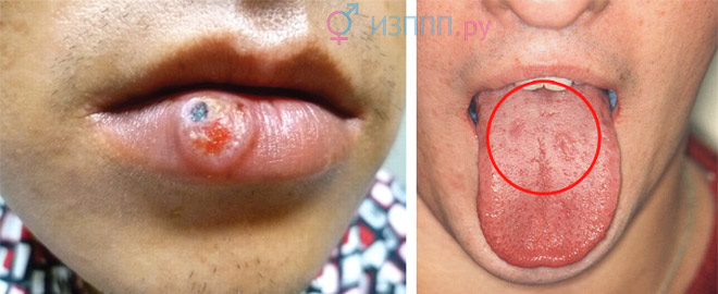
manifestations of syphilis on the lips (chancre) and tongue
Syphilides on the tongue, in the corners of the mouth due to constant irritation, they grow and rise above the mucous membranes and healthy skin, dense, the surface is grayish in color. They may become covered with erosions or ulcerate, causing pain. Papular syphilides on vocal cords At first they manifest themselves as hoarseness, later a complete loss of voice is possible - aphonia.
Syphilitic nail damage(onychia and paronychia): papules are localized under the bed and at the base of the nail, visible as reddish-brown spots. Then the nail plate above them becomes whitish and brittle, and begins to crumble. With purulent syphilide, severe pain is felt, the nail moves away from the bed. Subsequently, crater-shaped depressions form at the base, and the nail becomes three or four times thicker than normal.
Tertiary period of syphilis
Tertiary syphilis manifests itself as focal destruction of the mucous membranes and skin, any parenchymal or hollow organs, large joints, and nervous system. Main features – papular rashes and gummas, degrading with rough scarring. Tertiary syphilis is rarely detected and develops within 5-15 years if no treatment is provided. Asymptomatic period ( latent syphilis) can last more than two decades, diagnosed only by serological tests between secondary and tertiary syphilis.

what can affect advanced syphilis
Papular elements dense and round, up to 1 cm in size. They are located deep in the skin, which becomes bluish-red above the papules. Papules appear at different times and are grouped into arcs, rings, and elongated garlands. Typical for tertiary syphilis focus rash: each element is determined separately and in its own stage of development. The disintegration of papular syphilomas begins from the center of the tubercle: round ulcers appear, the edges are steep, there is necrosis at the bottom, and a dense ridge along the periphery. After healing, small dense scars with a pigment border remain.
Serpinginous Syphilide is grouped papules that are in different stages of development and spread over large areas of the skin. New formations appear along the periphery, merging with old ones, which at this time are already ulcerated and scarred. The sickle-shaped process seems to crawl towards healthy areas of the skin, leaving a trail of mosaic scars and foci of pigmentation. Numerous tuberculate compactions create a motley picture truly polymorphic rash, which is visible in the later periods of syphilis: different sizes, different morphological stages of the same elements - papules.

syphilitic gumma on the face
Syphilitic gumma. At first it is a dense node, which is located deep in the skin or under it, mobile, up to 1.5 cm in size, painless. After 2-4 weeks, the gumma is fixed in relation to the skin and rises above it as a rounded dark red tumor. Softening appears in the center, then a hole forms and the sticky mass comes out. In place of the gumma, a deep ulcer forms, which can increase along the periphery and spread along an arc ( serping gummous syphilide), and in the “old” areas healing occurs with the appearance of retracted scars, and in new areas – ulceration.
Most often, syphilitic gummas are located alone and are localized on the face, near the joints, and on the front of the legs. Closely located syphilides can merge to form gumm pad and turn into impressive ulcers with compacted, jagged edges. In weakened patients, when syphilis is combined with HIV, gonorrhea, viral hepatitis, gummas may grow in depth - mutilating or irradiating gummas. They disfigure the appearance and can even lead to the loss of an eye, testicle, perforation and death of the nose.
Gunma in the mouth and inside the nose disintegrate with destruction of the palate, tongue and nasal septum. Defects are formed: fistulas between the cavities of the nose and mouth (voice is nasal, food may enter the nose), narrowing of the throat opening(difficulty swallowing), cosmetic problems – failed saddle nose. Language At first it enlarges and becomes lumpy, after scarring it shrinks, and it becomes difficult for the patient to speak.
Visceral and neurosyphilis
At visceral In tertiary syphilis, organ damage is observed, with the development neurosyphilis– symptoms from the central nervous system (CNS). During the secondary period, early syphilis of the central nervous system appears; it affects the brain, its vessels and membranes ( meningitis And meningoencephalitis). In the tertiary period, manifestations of late neurosyphilis are observed, these include optic atrophy, tabes dorsalis and progressive paralysis.

Tabes dorsalis– manifestation of syphilis of the spinal cord: the patient literally does not feel the ground under his feet and cannot walk with his eyes closed.
Progressive paralysis maximum manifests itself one and a half to two decades after the onset of the disease. The main symptoms are mental disorders, from irritability and memory impairment to delusional states and dementia.
Optic atrophy: with syphilis, one side is first affected, and a little later vision deteriorates in the other eye.
Gummas affecting the head brain, are rarely observed. According to clinical signs, they are similar to tumors and are expressed by symptoms of brain compression - increased intracranial pressure, rare pulse, nausea and vomiting, prolonged headaches.

bone destruction due to syphilis
Among visceral forms it predominates syphilis of the heart and vascular system(up to 94% of cases). Syphilitic mesaortitis– inflammation of the muscular wall of the ascending and thoracic aorta. Often found in men, it is accompanied by dilation of the artery and symptoms of cerebral ischemia (dizziness and fainting after exercise).
Syphilis liver(6%) leads to the development of hepatitis and liver failure. The total proportion of syphilis of the stomach and intestines, kidneys, endocrine glands and lungs does not exceed 2%. Bones and joints: arthritis, osteomyelitis and osteoporosis, consequences of syphilis - irreversible deformities and blockade of joint mobility.
Congenital syphilis
Syphilis can be transmitted during pregnancy, from an infected mother to her child at 10-16 weeks. Frequent complications are spontaneous abortions and fetal death before birth. Based on time criteria and symptoms, congenital syphilis is divided into early and late.
Early congenital syphilis
 Children with obvious underweight, with wrinkled and sagging skin, resemble little old people. Deformation of the skull and its facial part (“Olympic forehead”) is often combined with dropsy of the brain and meningitis. Present keratitis– inflammation of the cornea of the eyes, loss of eyelashes and eyebrows is visible. Children aged 1-2 years develop syphilitic rash, localized around the genitals, anus, on the face and mucous membranes of the throat, mouth, nose. The healing rash forms scarring: scars that look like white rays around the mouth are a sign of congenital lues.
Syphilitic pemphigus– a rash of vesicles, observed in a newborn several hours or days after birth. It is localized on the palms, skin of the feet, on the folds of the forearms - from the hands to the elbows, on the torso.
Rhinitis, the causes of its occurrence are syphilides of the nasal mucosa. Small purulent discharge appears, forming crusts around the nostrils. Breathing through the nose becomes problematic, the child is forced to breathe only through the mouth.
Osteochondritis, periostitis– inflammation and destruction of bones, periosteum, cartilage. Most often found on the legs and arms. Local swelling, pain and muscle tension are noted; then paralysis develops. During early congenital syphilis, destruction of the skeletal system is diagnosed in 80% of cases.
Late congenital syphilis
Late form manifests itself in the age period of 10-16 years. The main symptoms are weakened vision with the possible development of complete blindness, inflammation of the inner ear (labyrinthitis) followed by deafness. Skin and visceral gummas are complicated by functional disorders of organs and disfiguring scars. Deformation of teeth and bones: the edges of the upper incisors have semilunar notches, the shins are curved, and due to the destruction of the septum, the nose is deformed (saddle-shaped). Problems with the endocrine system are common. The main manifestations of neurosyphilis are tabes dorsalis, epilepsy, speech impairment, progressive paralysis.
Congenital syphilis is characterized by a triad of symptoms Hutchinson:
- teeth with an arched edge;
- cloudy cornea and photophobia;
- labyrinthitis – tinnitus, loss of orientation in space, weakened hearing.
How is syphilis diagnosed?
Diagnosis of syphilis is based on clinical manifestations characteristic of different forms and stages of the disease, and laboratory tests. Blood taken to conduct a serological (serum) test for syphilis. To neutralize teponems, specific proteins are produced in the human body - which are determined in the blood serum of someone infected or sick with syphilis.
RW analysis blood (Wassermann reaction) is considered obsolete. It can often be false-positive for tuberculosis, tumors, malaria, systemic diseases and viral infections. Among women– after childbirth, during pregnancy, menstruation. Consuming alcohol, fatty foods, and certain medications before donating blood for RW may also cause unreliable interpretation of the syphilis test.
Based on the ability of antibodies (immunoglobulins IgM and IgG) present in the blood of people infected with syphilis to interact with antigen proteins. If the reaction has passed, analysis positive, that is, the causative agents of syphilis were found in the body of a given person. Negative ELISA – there are no antibodies to treponema, there is no disease or infection.

The method is highly sensitive, applicable for the diagnosis of latent - hidden forms - syphilis and checking people who had contact with the patient. Positive even before the first signs of syphilis appear (by IgM - from the end of the incubation period), and can be determined after the complete disappearance of treponemes from the body (by IgG). ELISA for the VRDL antigen, which appears during alteration (“deterioration”) of cells due to syphilis, is used to monitor the effectiveness of treatment regimens.
RPHA (passive hemagglutination reaction)– gluing of red blood cells that have antigens on their surface Treponema pallidum, with specific antibody proteins. RPHA is positive in case of illness or infection with syphilis. Remains positive throughout the patient’s life, even after full recovery. To exclude a false-positive response, RPGA is supplemented with ELISA and PCR tests.
Direct methods laboratory tests help identify the causative microorganism, and not antibodies to it. Using this, you can determine the DNA of treponemes in biomaterial. Microscopy smear from the serous discharge of a syphilitic rash - a method for visual detection of treponemes.
Treatment and prevention
Treatment of syphilis is carried out taking into account the clinical stages of the disease and the patient's susceptibility to drugs. Seronegative early syphilis is easier to treat; in late variants of the disease, even the most modern therapy is not able to eliminate consequences of syphilis– scars, organ dysfunction, bone deformities and nervous system disorders.

There are two main methods of treating syphilis: continuous(permanent) and intermittent(course). During the process, control tests of urine and blood are required; the well-being of patients and the functioning of organ systems are monitored. Preference is given to complex therapy, which includes:
- Antibiotics(specific treatment of syphilis);
- General strengthening(immunomodulators, proteolytic enzymes, vitamin-mineral complexes);
- Symptomatic drugs (painkillers, anti-inflammatory, hepatoprotectors).
Prescribe a diet with an increased proportion of complete proteins and a limited amount of fat, and reduce physical activity. Sexual contact, smoking and alcohol are prohibited.
Psychological trauma, stress and insomnia negatively affect the treatment of syphilis.
Patients with early latent and contagious syphilis undergo the first course of 14–25 days in the clinic, then are treated on an outpatient basis. Treatment for syphilis begins with penicillin antibiotics– sodium or potassium salt of benzylpenicillin, bicillins 1-5, phenoxymethylpenicillin are administered intramuscularly. A single dose is calculated based on the patient’s weight; if there are inflammatory signs in the cerebrospinal fluid (spinal fluid), then the dosage is increased by 20%. The duration of the entire course is determined according to the stage and severity of the disease.
Permanent method: the starting course for seronegative primary syphilis will require 40-68 days; seropositive 76-125; secondary fresh syphilis 100-157.
Course treatment: tetracyclines are added to penicillins ( doxycycline) or macrolides ( azithromycin), bismuth-based preparations – bismovrol, bijoquinol, and iodine - potassium or sodium iodide, calcium iodine. Cyanocobalamin (Vit. B-12) and solution koamida enhance the effect of penicillin and help increase the concentration of the antibiotic in the blood. Injections of pyrogenal or prodigiosan, autohemotherapy, and aloe are used as nonspecific therapy for syphilis, increasing resistance to infection.
During pregnancy, syphilis is treated only with penicillin antibiotics, without drugs with bismuth salts.
Proactive(preventive) treatment: carried out as in the case of seronegative primary syphilis, if sexual contact with an infected person was 2-16 weeks ago. One course of penicillin is used for drug prevention of syphilis if contact occurred no more than 2 weeks ago.
Prevention of syphilis– identification of infected people and their circle of sexual partners, preventive treatment and personal hygiene after sexual intercourse. Examinations for syphilis of people belonging to risk groups - doctors, teachers, staff of kindergartens and catering establishments.
Video: syphilis in the program “Live Healthy!”
Video: syphilis in the STD encyclopedia
Description of the disease
The statement that syphilis is exclusively a disease that is sexually transmitted is not entirely true. The fact is that you can become infected with it in everyday life when the infection directly enters the bloodstream through scratches or wounds on the body; this is also possible when using toilet items (towel, washcloth) belonging to the patient.
In addition, infection with syphilis can occur through blood transfusion, and syphilis can also be congenital. Basically, the rash is located in the areas of hair and steps, as well as on the palms.
In addition, in women it is also localized under the mammary glands; for both sexes, its concentration can be located in the genital area.
After 3-4 weeks from the moment of infection, the place where Treponema pallidum, the causative agent of infection of this disease (which is mainly the genitals), was introduced acquires signs indicating primary syphilis.
Syphilitic rash is a modification of the superficial vessels of the skin. Treponema pallidum, entering the blood, releases specific toxins that dilate blood vessels. Further, the vascular reaction depends on the state of the immune system. Each person is individual, and so is their immune response.
Simple dilatation of blood vessels on the skin appears in the form of spots (roseola). Such spots easily disappear when pressed (the blood vessels are compressed and the skin becomes pale).
If there is an increase in the permeability of the vascular wall, plasma partially accumulates around the vessel along with immune cells, an inflammatory reaction occurs and a hard “muff” is formed around the dilated vessel.
On the skin this appears as a small rounded lump, i.e. a nodule (papule) is formed.
If the immune system is weakened, bacteria begin to actively multiply outside the vascular bed. The immune system, protecting the body, forms an inflammatory capsule around the largest accumulations of bacteria, inside which pus accumulates. This manifestation of the immune reaction on the skin looks like pustules (pustules).
Most people believe that you can only become infected with syphilis through sexual intercourse, and if a man or woman keeps their intimate relationships clean, they will not be at risk of this disease.
This opinion is erroneous, since transmission of infection is possible both through household contact and during medical procedures in dubious institutions where sterility conditions are not observed.
Direct blood transfusion, which is used in emergency cases, is also dangerous: the donor may not know about his illness, which will lead to infection of the recipient.
The third way is from an infected woman to her child.
Syphilis is a classic venereal (i.e. sexually transmitted) disease, which affects males and females equally. Most people get sick with syphilis during their reproductive years: men from 16-18 to 65-70 years old, women from 16 to 35-45 years old.
Syphilis - what is it? Syphilis is a serious disease, which is characterized by the pathological process affecting the skin, mucous membranes and internal organs of the patient.
The causative agent of syphilis is a microorganism called spirochete pallidum. It looks like a curved spiral, can move in different ways, and can divide transversely.
Favorable conditions for the development of this bacterium are found in the human lymphatic tract and nodes, so it is there that it begins to rapidly multiply. The presence of such microorganisms in the blood can be detected at the stage of the secondary type of disease.
Bacteria can remain in a warm and humid environment for quite a long time; the most optimal temperature is 37°C. In addition, they are resistant to low temperatures.
Pathogenic microorganisms die when dried, heated to 55°C-100°C, or treated with disinfectants, acidic or alkaline solutions.
Household syphilis, symptoms and treatment, prevention, photo can lead to many negative consequences for human health, even ending very tragically. But the prognosis depends on whether this dangerous disease is detected in a timely manner.
Types of rash with syphilis
After the disappearance of primary hard chancre and the development of the secondary stage, new rashes begin to cover the body. The rash on the body with secondary syphilis is very diverse
- Roseola are pale pink spots that most often cover the abdomen and side of the patient’s torso. They do not have clear contours, do not merge, and do not cause discomfort. Roseola is considered the most common type of rash, as it is observed in 90% of Lewis patients.
- Papules are round nodules, no larger than a pea. The first days after formation are smooth, but after that they may peel off. The papular rash of syphilis is usually observed on the palms, soles, anus and genitals.
- Palmoplantar syphilides is another type of papules, characterized by clear contours and a typical color - bright red or purple. It mainly affects the palms and soles of the feet. Sometimes they are confused with calluses, so people put off visiting the doctor. A few days after formation, they crack and begin to peel off.
There are such types of rashes with syphilis:
- First stage. The manifestation of this stage can be noticed a month after the infection was introduced into the body. At this moment, the first signs of syphilis can be observed. The rash appears as red pimples, which after a certain time take on the appearance of ulcers. The rash may disappear after a couple of weeks, but will soon reappear. Such a rash can remain on the human body for a long time, even be present for several years.
Stages of the disease
There are several stages through which patients infected with syphilis go:
Signs of primary syphilis include the appearance of a small red spot that turns into a lump after a few days. The center of the tubercle is characterized by gradual necrosis of the tissue (its death), which ultimately forms a painless ulcer framed by hard edges, that is, chancre.
The duration of the primary period is about seven weeks, after the start of which, after about a week, all lymph nodes undergo enlargement.

The completion of the primary period is characterized by the formation of many pale treponemas, causing treponemal sepsis. The latter is characterized by weakness, general malaise, joint pain, fever and, in fact, the formation of a characteristic rash, which indicates the onset of the secondary period.
The secondary stage of syphilis is extremely diverse in its symptoms and it is for this reason that in the 19th century French syphilidologists called it the “great ape”, thereby indicating the similarity of the disease at this stage with other types of skin diseases.
Signs of the general type of secondary stage of syphilis include the following features of the rash:
- Absence of subjective sensations (pain, itching);
- Dark red color of the rash;
- Density;
- Clarity and regularity of roundness or roundness of outlines without their tendency to possibly merge;
- Peeling of the surface is of an unexpressed nature (in most cases its absence is noted);
- Spontaneous disappearance of formations is possible without subsequent atrophy and scarring.
Most often, rashes of the secondary stage of syphilis are characterized by the following manifestations (see photo of a syphilitic rash):
This stage of the disease is characterized by a small amount of Treponema pallidum in the body, but it is sensitized to their effects (that is, allergic).
This circumstance leads to the fact that even with the influence of a small amount of treponemes, the body responds with a peculiar form of anaphylactic reaction, which consists in the formation of tertiary syphilides (gummas and tubercles).
Their subsequent breakdown occurs in such a way that characteristic scars remain on the skin. The duration of this stage can be decades, which ends with deep damage to the nervous system.
Dwelling on the rash of this stage, we note that the tubercles are smaller in size when compared with gummas, both in their size and in the depth at which they occur.
Tubercular syphilis is determined by palpating the thickness of the skin and identifying a dense formation in it. It has a hemispherical surface, the diameter is about 0.3-1 cm.
Above the tubercle, the skin becomes bluish-reddish in color. The tubercles appear at different times, grouping into rings.


- Isolating the contour of a liquid drop in the problem of determining surface tension Isolating the contour of a drop
- Method for producing calcium peroxide
- Feo color. Chemical properties. Refractory black pyrophoric powder, insoluble in water. Methods for obtaining iron
- Sodium thiosulfate Natrii thiosulfas (ln) Thiosulfate chemical formula
- Rhodanide method for determining iron
- Baking soda: formula, application
- Carbonates. Baking soda formula. Baking soda: formula, application Safety of baking soda for humans
- Description of the disease, signs, treatment
- Liver diseases: causes, types, symptoms and prevention What is a virus in the liver
- Helminthic infestation: causes, symptoms, treatment, prevention
- Folk remedies
- Food fermentation and its importance
- Study of pharmacokinetics and bioavailability - journal of pharmacokinetics and pharmacodynamics High performance liquid chromatography
- Test work chloroform Chloroform at home
- Noble gases and their properties Noble gases what causes the inertness of these gases
- Raman and NIR spectroscopy Problems of NIR spectrometry and how to solve them
- Side specialties of tantalum
- What is tantalum and where is it used?
- Molecular effects of enzyme action
- Cervical dysplasia: first signs, symptoms and treatment