Directions of evolution of gill type respiration. respiration in animals. Features of respiration in animals of different groups Characteristic features of the respiratory organs
Evolution of the respiratory system
Stages of the breathing process
Breath- a set of processes that ensure the supply of oxygen from the environment to the body, which is necessary for the oxidation of organic substances in the mitochondria of the cell, and the release of carbon dioxide
Breath types:

Breath type:
Cellular.
Organisms: unicellular animals (amoeba, green euglena, infusoria slipper); coelenterates (jellyfish, coral polyps); some worms.
Single-celled organisms absorb oxygen dissolved in water over the entire surface of the body by diffusion.
Oxygen is involved in the breakdown of complex organic substances, resulting in the release of energy, which is necessary for the life of the animal.
Carbon dioxide formed as a result of respiration is also released outward through the entire surface of the body.
Tracheal breathing is breathing with the help of a system of combined tracheal tubes that permeate the entire body.
Organisms: class Insects (beetles, butterflies, grasshoppers, flies)
The abdomen of an insect is divided into 5–11 parts (segments). Each of them has a pair of small holes - spiracle. From each spiracle branching tubules extend inward - trachea that permeate the entire body of the insect. Watching the cockchafer, you can see how its abdomen either decreases in volume or increases. These are breathing movements. When you inhale, air containing oxygen enters the body through the spiracles, and when you exhale, air saturated with carbon dioxide exits.
In spiders (class Arachnids), the respiratory organs are represented not only by tracheae, but also by lung sacs that communicate with the external environment through respiratory openings.
Gill breathing is breathing with the help of specialized formations with a dense network of blood vessels.
Organisms: many aquatic life (fish, crayfish, molluscs)
Fish breathe oxygen dissolved in water with the help of special branched skin outgrowths called gills.
Fish are constantly swallowing water. From the oral cavity, water passes through the gill slits, wash the gills and exits from under the gill covers. Gills consist of gill arches and gill filaments which are pierced by many blood vessels. From the water that washes the gills, oxygen enters the blood, and carbon dioxide is removed from the blood into the water. The gills inside the body are called internal gills.
Some animals, such as amphibians, have thick tufts of gills on the surface of the body. Such gills are called - outdoor. Such is the structure of Proteus, a blind cave animal from the western regions of Yugoslavia, and axolotls (which are similar in general appearance to newts) - their homeland is Mexico.
The gill apparatus in chordates evolved towards the formation of gill filaments. In particular, 4-7 gill sacs have developed in fish, which are gaps between the gill arches and contain a large number of petals that are pierced by capillaries (Fig. 190). In fish, an air bladder also participates in respiration.[ ...]
Gill respiration is a typical aquatic respiration. The physiological purpose of the gills is to supply the body with oxygen. They transfer oxygen from the external environment to the blood.[ ...]
Skin respiration, as the most primary in phylogenesis and ontogenesis, is then replaced by a special, gill respiration, but still continues to play a certain role until the end of the life of the fish.[ ...]
Respiratory system. The gills are the organs of respiration. They lie on both sides of the head. They are based on gill arches. In the vast majority of cases, in our freshwater fishes, with the exception of lampreys only, the gills are covered on the outside with covers, and their cavity communicates with the oral cavity. On the gill arches there are two-row gill plates. Each gill plate is oblong, pointed, tongue-shaped, has a cartilaginous stamen at its base, enclosed in a bone cap and reaching to its free end. Along the inner edge of the gill plate runs a branch of the branchial artery, which brings venous blood, and along the outer edge - a branch of the branchial vein, which drains arterial blood. Hair vessels depart from them. On both flat sides of the gill plate are leaf-like plates, which actually serve for respiration or gas exchange. If there is only one row of plates on the gill arch, then it is called a semi-gill.[ ...]
In gobies, breathing in moist air is provided by the scalp, oral and gill cavities. The mucous membrane of these cavities is well supplied with blood vessels. Air is taken in by the mouth, oxygen is taken up in the mouth or gill cavity, and the remaining gas is expelled back through the mouth. Interestingly, many gobies do not have a swim bladder, and other organs are adapted for air breathing.[ ...]
In a number of fish, gill respiration in the early stages of development does not fully satisfy the needs of the organism. As a result, additional organs develop (subintestinal, superior tail and dorsal veins), which serve as a significant addition to gill breathing. With the development and improvement of gill respiration, embryonic respiration is gradually reduced.[ ...]
In addition to the frequency of respiration, changes are also observed in the depth of respiration. Fish in some cases (with low P02, elevated temperature, high CO2 content in the water) breathe very often. The respiratory movements themselves are small. Such shallow breathing is especially easy to observe at elevated temperatures. In some cases, the fish takes a deep breath. Mouth and gill covers open and close wide. With shallow breathing, the respiratory rhythm is large, with deep breathing it is small.[ ...]
Observing the rhythm of fish breathing, M. M. Voskoboinikov came to the conclusion that the passage of water in one direction through the mouth, gill filaments and gill openings is ensured by the work of the gill covers and the special position of the gill filaments.[ ...]
As the gill type of breathing develops, salmon use oxygen more easily, even if the latter is in a low concentration (decrease in the threshold concentration of O2).[ ...]
The ratio of main respiration to additional respiration varies in different fish. Even in the loach, intestinal respiration has changed from supplementary to almost equal to gill respiration. Vyun still needs. intestinal respiration, even if it is in well-aerated water. From time to time, it rises to the surface and swallows air, and then sinks to the bottom again. If, for example, in a perch or carp, with a lack of oxygen, the respiratory rhythm quickens, then the loach in. under such conditions, it does not speed up the rhythm of breathing, but uses intestinal respiration more intensively.[ ...]
Water is pumped through the gill cavity by the movement of the mouthparts and gill covers. Therefore, the respiratory rate in fish is determined by the number of movements of the gill covers. The breathing rhythm of fish is primarily affected by the oxygen content in the water, as well as the concentration of carbon dioxide, temperature, pH, etc. Moreover, the sensitivity of fish to a lack of oxygen (in water and blood) is much higher than to an excess of carbon dioxide (hypercapnia) . For example, at 10 ° C and a normal oxygen content (4.0-5.0 mg / l), trout performs 60-70, carp - 30-40 respiratory movements per minute, and at 1.2 mg 02 / l, the respiratory rate increases 2-3 times. In winter, the carp's breathing rhythm slows down sharply (up to 3-4 breaths per minute).[ ...]
With the mouth open and the gill covers closed, the zoda enters the oral cavity, passes between the gill filaments into the gill cavity. This is a breath. Then the mouth closes, and the operculum opens slightly and the water comes out. This is exhalation. Consideration of this process in detail led to two different ideas about the mechanism of respiration.[ ...]
In some fish, the pharynx and gill cavity are adapted for air breathing.[ ...]
Gills are the main respiratory organ in most fish. However, examples can be given where in some fish the role of gill respiration is reduced, while the role of other organs in the process of respiration is increased. Therefore, it is not always possible to answer the question of what the fish breathes at the moment. Having significantly expanded Bethe's table, we present the ratios of different forms of respiration in fish under normal conditions (Table 85).[ ...]
The inhibitory effect of excess CO2 on gill respiration and the stimulation of pulmonary respiration in lungfish have been repeatedly noted. The transition of lungfish from water to air breathing is accompanied by a decrease in arterial p02 and an increase in pCO2. It should be especially noted that the stimulation of air respiration and the suppression of water respiration in lungfish occurs under the influence of a decrease in the level of 02 in the water and an increase in the level of CO2. True, during hypoxia in lungfish ((Cheosegagoskk) both pulmonary and gill respiration intensifies, and during hypercapnia, only pulmonary respiration. It is curious that under the combined action of hypoxia and hypercapnia, ventilation of the lungs increases, and gills decrease. in the region of the gills or in the efferent gill vessels.[ ...]
The underdevelopment or complete absence of the gill cover makes it difficult to breathe and leads to disease of the gills. The slanting snout interferes with food intake. The arched back and pug-shaped head lead to a significant stunting.[ ...]
The most common type of intestinal respiration is that in which air is driven through the intestines and gas exchange occurs in the middle or back part of it (loaches, some catfishes). In another type, such as Hippostomos and Acarys, the air, after being in the intestines for some time, does not exit through the anus, but is squeezed back into the oral cavity and then thrown out through the gill slits. This type of intestinal breathing is fundamentally different from the first; subsequently, in some fish, it developed into pulmonary respiration.[ ...]
A more complex adaptation for air breathing is the supra-gill organ. The nadzhaberny organ is available in Opy-ocephalus (snakehead), living in the river. Cupid, in Luciocephalus, in Anabas, etc. This organ is formed by a protrusion of the pharynx, and not the gill cavity proper, as in labyrinth fish.[ ...]
Respiratory movements, respiratory rhythm. In fish, the gill cover periodically opens and closes. These rhythmic movements of the operculum have long been known as respiratory movements. However, a correct understanding of the breathing process has been achieved relatively recently.[ ...]
It is quite obvious that the intensity of skin respiration is an expression of the adaptability of fish to life in conditions of oxygen deficiency, when gill respiration is not able to provide the body with oxygen in the required amount.[ ...]
A general rule is observed: with the development of air respiration, the gill respiration decreases (Suvorov). Anatomically, this is expressed in the shortening of the gill filaments (in Polypterus, Ophiocephalus, Arapaima, Electrophorus) or in the disappearance of a number of lobes (in Monopterus, Amphipnous and lungfish). In protopterus, for example, petals are almost completely absent on the first and second arches, while in lepidosiren, gill filaments are poorly developed.[ ...]
The fish of warm waters have a device for air breathing in the form of a labyrinth. The labyrinth organ is formed by a protrusion of the gill cavity proper and is sometimes (as in Anatas) supplied with its own musculature. The inner surface of the “labyrinth cavity” has a variety of curvatures due to curved bone plates covered with a mucous membrane. Many blood vessels and capillaries approach the surface of the "labyrinth cavity". Blood enters them from a branch of the fourth afferent branchial artery. Oxygenated blood flows into the dorsal aorta. The air captured by the fish in the mouth enters the labyrinth from the oral cavity and gives oxygen to the blood there.[ ...]
More recently, S. V. Streltsova (1949) has carried out more detailed studies of skin respiration in 15 species of fish. It determined both general respiration and specifically skin respiration. Gill respiration was turned off by applying a hermetic rubber mask to the gills. This technique allowed her to determine the share of skin respiration in the total respiration of fish. It turned out that this value is very different for different fish and is associated with the way of life and ecology of fish.[ ...]
Experiments have shown that the V, VII, IX and X pairs of head nerves are necessary for normal breathing. Branches from them innervate the upper jaw (V pair), gill cover (VII pair) and gills (IX and X pairs).[ ...]
In practice, all cyclostomes and fish have a "morphofunctional reserve" for increasing the power of respiration in the form of some "higher" gas exchange structures. It has been experimentally established that no more than 60% of the gill filaments function in fish under normal conditions. The rest are switched on only in conditions of impending hypoxia or with an increase in oxygen demand, for example, with an increase in swimming speed.[ ...]
In the larval stage (tadpoles), amphibians are very similar to fish: they retain gill breathing, have fins, a two-chambered heart, and one circle of blood circulation. Adult forms are characterized by a three-chambered heart, two circles of blood circulation, two pairs of limbs. The lungs appear, but they are poorly developed, so additional gas exchange occurs through the skin (Fig. 81). Amphibians live in warm, humid places, and are especially common in the tropics, where they are most numerous.[ ...]
Sturgeon larvae and fry are transported in the first two days after hatching from eggs before switching to gill breathing, since gill breathing requires more oxygen. Saturation of water with oxygen should be at least 30% of normal saturation. At a water temperature of 14-17 ° C and constant aeration, the planting density, depending on the mass of larvae, can be increased to 200 pcs. per 1 liter of water.[ ...]
At the age of 15 days, the larva has enlarged axillary veins that encircle the intestines (already performing the function of respiration), and a pectoral fin with densely branched vessels. At the age of 57 days, the external gills of the larva have shrunk and are completely covered by the gill cover. All. fins, except preanal, well supplied with vessels. These fins serve as respiratory organs (ri £.-67).[ ...]
In a carefully performed test on the same species of fish - on brook trout, it was shown that already at pH 5.2, hypertrophy of the mucous cells of the gill epithelium occurs, and mucus accumulates on the gills. Subsequently, with an increase in water acidity to 3.5, destruction of the gill epithelium and its rejection from supporting cells was noted. The accumulation of mucus on the gills during the period when breathing is especially difficult has also been noted in other salmon species.[ ...]
It is necessary to increase the pO2 at which HbO2 is formed. For the most part, gill breathing and heart rate increase in fish. In this case, not only the maintenance of p02 at a higher level occurs, but also a decrease in pCO2. However, the body can only achieve this within certain temperature limits, since water in a reservoir is less oxygenated at elevated temperatures than at low temperatures. Under laboratory conditions and when transporting live fish in closed vessels, the condition of the fish can be improved by: that with an increase in temperature, the RH in the water is increased artificially, by aeration.[ ...]
The supragillary and labyrinth organs are found in the snakehead and in tropical fish (cockerel, gourami, macropods). They are sac-like protrusions of the gill cavity (labyrinth organ) or pharynx (supra-gill organ) and are intended mainly for air breathing.[ ...]
In the European bitterling, the vessels of the respiratory network reach a greater development than in our other cyprinids. This is the result of the adaptation of the organism to life in the gill cavity of mollusks in the early stages of development under poor oxygen conditions. With the transition to life in water, all these adaptations disappear and only developed gill breathing remains.[ ...]
Fish are divided into cartilaginous and bony. The habitat of fish is water bodies, which shaped the features of their body and created fins as organs of movement. Breathing is gill, and the heart is two-chambered and one circle of blood circulation.[ ...]
According to R. Lloyd, the leading moment in this case is an increase in the flow of water passing through the gills, and, as a result, an increase in the amount of poison reaching the surface of the gill epithelium with subsequent penetration into the body. Moreover, the concentration of poison on the surface of the gill epithelium is determined not only by the concentration of the poison in the bulk of the solution, but also by the rate of respiration. We add to this that according to the data obtained by M. Shepard, with a decrease in the concentration of oxygen in the water, the content of hemoglobin in the blood increases and, most importantly, the rate of blood circulation through the gills increases.[ ...]
By the way, the same ability was used to explain the cases of Kgrp life with overgrown mouths. And here, studies have shown that these carps drag out their existence for some time, having adapted to take in water for breathing and, along with it, a certain number of crustaceans through the gill openings.[ ...]
Chordates are also characterized by the presence of a nerve bundle in the form of a tube above the notochord and a digestive tube under the notochord. Further, they are characterized by the presence in the embryonic state or throughout life of numerous gill slits that open outward from the pharyngeal region of the digestive tube and are respiratory organs. Finally, they are characterized by the location of the heart or its replacement vessel on the ventral side.[ ...]
Summarizing the numerous experimental data available today on the effect of long-term or short-term oxygen deficiency on fish of various ecologies, a number of general conclusions can be drawn. The primary reaction of fish to hypoxia is an increase in respiration by increasing its frequency or depth. The volume of gill ventilation at the same time increases sharply. The heart rate drops, the stroke volume of the heart increases, as a result of which the volume of blood flow remains constant. During the development of hypoxia, oxygen consumption initially increases slightly, then returns to normal. As hypoxia deepens, the efficiency of oxygen absorption begins to decrease, while oxygen consumption by tissues increases, which creates additional difficulties for fish in providing oxygen demand under conditions of its low content in water. Oxygen tension in arterial and venous blood, utilization of oxygen from water, the efficiency of its transfer and the efficiency of blood oxygenation are reduced.[ ...]
The recording of the electrocardiogram is carried out as follows. Electrodes soldered onto thin flexible conductors are inserted: one into the region of the heart on the ventral side of the body, and the other - between the dorsal fin and the head on the dorsal side. To record the respiratory rate, electrodes are inserted into the operculum and into the rostrum. Respiratory rate and heart rate can be recorded simultaneously through two independent channels of an electrocardiograph or any other device (for example, a two-channel electroencephalograph). In this case, the fish can be both in a free state in an aquarium, and in a fixed one. Recording an electrocardiogram is feasible only in conditions of complete screening of the aquarium water. Screening can be done in two ways: by immersing a plate of galvanized iron in water or by soldering a conductor to the bottom of the aquarium. If the aquarium is plexiglass, it should be installed on a sheet of iron.[ ...]
Comparing these data for juveniles with those of Kuptsis for adult roach, it is easy to see that the threshold value in juvenile roach on the 49th day after hatching is very close to the threshold value for the adult (1 and 0.6-1 mg/l, respectively). Consequently, after the establishment of gill respiration, the ability to use oxygen quickly reaches its limit.[ ...]
The gills play an important role in removing excess salts. If divalent ions are excreted in significant quantities through the kidneys and the digestive tract, then monovalent ions (mainly Na and CG) are excreted almost exclusively through the gills, which in fish perform a dual function - respiration and excretion. In the gill epithelium there are special large goblet cells containing a large number of mitochondria and a well-developed eudoplasmic reticulum. These "chloride" (or "salt") cells are located in the primary gill filaments and, unlike the respiratory cells, are associated with the vessels of the venous system. The transfer of ions through the gill epithelium has the character of active transport and proceeds with the expenditure of energy. The stimulus for the excretory activity of chloride cells is an increase in blood osmolarity.[ ...]
Suspended solids tend to form unstable or stable suspensions and include both inorganic and organic components. With an increase in their content, light transmission deteriorates, photosynthesis activity decreases, the appearance of water deteriorates, and gill respiration may be disturbed. As solid particles settle to the bottom, the activity of benthic flora and fauna decreases.[ ...]
In the ontogenesis of fish, a certain sequence of the role of individual oxygen-receiving surfaces is observed: the stellate sturgeon egg breathes the entire surface; in the embryo, oxygen supply occurs mainly through a dense network of capillaries on the yolk sac; after hatching, approximately on the 5th day, gill breathing appears, which then becomes the main one.[ ...]
The loach rises to the surface of the water to swallow air at: t = 10 ° 2-3 times per hour, and at 25-30 ° already 19 times. If the water is boiled, i.e., P02 is reduced, then the loach rises to the surface at t \u003d 25-2.7 ° 'once an hour. At t=5° in running water, it did not rise to the surface for 8 hours. These experiments show quite clearly that intestinal respiration, which is a supplement to gill respiration, copes quite satisfactorily with its function at low demands of the organism at 02 (at t = 5°) or at a high concentration of oxygen in the environment (running water). But gill respiration is not enough if the metabolism in the body is increased (t == 25-30°) or if the P02 in the medium (boiled water) is greatly reduced. In this case, intestinal respiration is additionally switched on, and the loach receives the required amount of oxygen.[ ...]
In the Devonian, the climate was sharply continental, arid, with sharp fluctuations in temperature during the day and seasons, extensive deserts and semi-deserts appeared. The first glaciations were also observed. During this period, fish reached a great prosperity, inhabiting the seas and fresh waters. At that time, many surface water bodies dried up in the summer, froze through, and the fish that inhabited them could be saved in two ways: burrowing into the silt or migrating in search of water. Lungfish took the first path, in which, along with gill breathing, pulmonary respiration developed (the lung developed from the swim bladder). Their fins looked like blades, consisting of individual bones with muscles attached to them. With the help of fins, fish could crawl along the bottom. In addition, they too could have pulmonary respiration. The lobe-finned fish gave rise to the first amphibians - stegocephals. On land in the Devonian, the first forests of giant ferns, horsetails and club mosses appear.[ ...]
Of the general clinical changes in fish, the following are noted: depression of the general condition, suppression and perversion of reactions to: external stimuli; darkening, pallor, hyperemia and hemorrhages on the skin of the body; ruffling of scales; violation of the sense of balance, orientation, coordination of movements and coordinated work of the fins; conjunctivitis, keratitis, cataracts, corneal ulceration, bulging eyes, loss of vision; complete or partial refusal to take food; swelling of the abdomen (acute cases of poisoning); change in the rhythm of breathing and the amplitude of oscillation of the gill covers; periodic cramps of the muscles of the body, tremor of the gill covers and pectoral fins. With chronic intoxication, signs of increasing exhaustion develop. In severe processes develops: toxic dropsy. In case of death, poisoned fish: sink from the surface of the water to the bottom, they develop a coma, breathing becomes shallow, then stops - death occurs.[ ...]
Less clear is the localization of peripheral receptors that perceive changes in CO2 content and the pathways for conducting impulses from these receptors to the respiratory center. So, for example, after transection of the IX and X pairs of cranial nerves innervating the gills, the impulses remained in a weakened form. In lung-breathing fish, gill respiration is noted to be inhibited with an increase in pCO2 in the water, which can be relieved by atropine. The effect of oppression of pulmonary respiration under the influence of excess carbon dioxide was not noted in these fish, which suggests the presence of CO2-sensitive receptors in the gill area.
Animal breath – set of processes that providehit into the body from the environmentoxygen , hiscell use for the oxidation of organic substances andbreeding from the body of carbon dioxide.This breathing is calledaerobic , and the organismsaerobes .
OK. No. 28. Biology.
green algae chlorella

Infusoria shoe

The process of respiration in animals is conditionally divided into three stages :
External respiration = gas exchange. Through this process, the animal receives oxygen and gets rid of carbon dioxide, which is the end product of metabolism.
Transport of gases in the body- this process is provided either by special tracheal tubes or internal body fluids (blood containing hemoglobin- a pigment that can attach oxygen and transport it to cells, as well as carry carbon dioxide out of cells).
internal breathing- occurs in cells. Simple nutrients (amino acids, fatty acids, simple carbohydrates) are oxidized and broken down with the help of cell enzymes, during which the ENERGY necessary for the life of the body is released.
The main significance of respiration is the release of energy from nutrients with the help of oxygen, which takes part in oxidation reactions.
Some of the simplest anaerobic organisms, i.e. organisms, not requiring oxygen.
Anaerobes are optional and obligatory. Facultative anaerobic organisms are organisms that can live both in the absence of oxygen and in its presence. Obligate anaerobic organisms are organisms for which oxygen is poisonous. They can only live in the absence of oxygen. Anaerobic organisms do not need oxygen to oxidize nutrients. 
Brachonella - anaerobic infusoria
Intestinal Giardia


human roundworm
By way of breathing and the structure of the respiratory apparatus in animals, 4 types of breathing are distinguished:
Skin respiration
is the exchange of oxygen and carbon dioxide through the integument of the body. This process is based on the most important physical process - diffusion
. Gases enter only in a dissolved state through the covers shallowly and at a low speed. Such breathing in organisms that are small in size, have wet covers, lead an aquatic lifestyle. This is - sponges, coelenterates, worms, amphibians.


Tracheal breathing
– 
carried out with the help
connected systems
tubules - trachea , which
permeate the entire body
participation of fluids. With
their environment
connect special
holes - spiracles.
Organisms with tracheal
breathing is also small in size (no more than 2 cm, otherwise the body will not have enough oxygen). This is - insects, centipedes, arachnids.
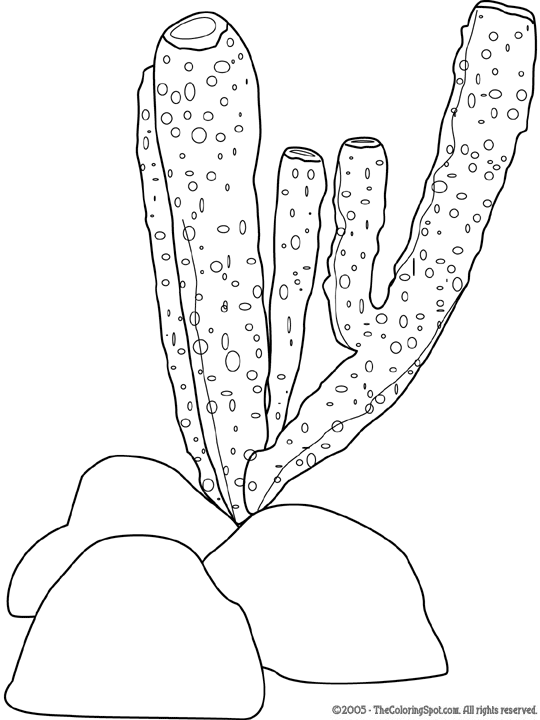
gill breathing - with the help of specialized formations with a dense network of blood vessels. These outgrowths are called gills . In aquatic animals polychaete worms, crustaceans, mollusks, fish, certain amphibian species. Invertebrates usually have external gills, while chordates have internal gills. Gill-breathing animals have additional forms of breathing through the skin, intestines, mouth surface, swim bladder.
Polychaete with gills

Crustacean gills

Nudibranch mollusk

Pulmonary respiration - this is breathing with the help of internal specialized organs - lungs.
Lungs– these are hollow thin-walled bags, braided with a dense network of tiny blood vessels - capillaries. Diffusion of oxygen from the air into the capillaries occurs on the inner surface of the lungs. Accordingly, the larger this inner surface, the more actively diffusion takes place.
Almost all terrestrial vertebrates breathe with lungs. reptiles, birds, some terrestrial invertebrates - spiders, scorpions, pulmonary mollusks, and some aquatic animals - lungfish. Air enters the lungs through Airways.

Lungs of a mammal


reptile lung
Respiratory system of birds
Breathing in animals is determined by their way of life and is carried out with the help of integument, trachea, gills and lungs.
Respiratory system – a set of organs for carrying air or water that contain oxygen, and gas exchange between the body and the environment.
Respiratory organs develop as outgrowths of the outer integument or walls of the intestinal tract. The respiratory system includes the respiratory tract and gas exchange organs. Vertebrates Airways – nasal cavity, larynx, trachea, bronchi ; a respiratory system -lungs .
Comparative characteristics of the respiratory organs.
|
Group |
Characteristic features of the respiratory system |
|
Coelenterates |
Gas exchange across the entire surface of the body. There are no special respiratory organs. |
|
annelids |
External gills (polychaete worms) and entire body surface (oligochaete worms, leeches) |
|
shellfish |
Gills (bivalves, cephalopods) and lungs (gastropods) |
|
arthropods |
Gills (crustaceans), tracheae and lungs (arachnoids), tracheae (insects) |
|
Fish |
Gills. Additional organs for breathing: lungs (lungfish), parts of the oral cavity, pharynx, intestines, swim bladder |
|
Amphibians |
Lungs are cellular, gills (in larvae), skin (with a large number of vessels). Respiratory tract: nostrils, mouth, tracheo-laryngeal chamber |
|
reptiles |
Light honeycomb. Respiratory tract: nostrils, larynx, trachea, bronchi |
|
Birds |
Light spongy. Respiratory tract: nostrils, nasal cavity, upper larynx, trachea, lower larynx with vocal apparatus, bronchi. There are air bags. |
|
mammals |
Light alveolar. Respiratory tract: nostrils, nasal cavity, larynx with vocal apparatus, trachea, bronchi. |
Functions of the respiratory system:
Delivery of oxygen to the cells of the body and removal of carbon dioxide from the cells of the body and gas exchange(main function).
Body temperature regulation(since water can evaporate through the surface of the lungs and airways)
Purification and disinfection of incoming air(nasal mucus)
Questions for self-control.
|
Grade |
Questions for self-control |
|
1. What is breathing? 2. The main stages of breathing? 3. Name the main types of animal breathing. 4. Give examples of animals that breathe with their skin, gills, windpipes and lungs. 5. What is the respiratory system? 6. Name the main functions of the respiratory system. |
|
|
7. What is the importance of breathing for the release of energy in animal cells? 8. What determines the type of breathing of animals? 9. What are the functions of the respiratory system? |
|
|
10. Describe how vertebrates breathe. |
Comparative characteristics of the respiratory organs of animals.
|
Respiratory system |
Structural features |
Functions |
Examples |
|
Gills
|
External(comb, filamentous and pinnate) or internal(always associated with the pharynx) thin-walled outgrowths of the body that contain many blood vessels |
Gas exchange in the aquatic environment |
In fish, in almost all larvae of anurans, in most molluscs, some worms and arthropods |
|
Trachea
|
Branched tubules that permeate the entire body and open outward with holes (stigmas) |
Gas exchange in the air |
In most arthropods |
|
Lungs
|
Thin-walled bags that have an extensive network of vessels |
Gas exchange in the air |
In some molluscs and fish, terrestrial vertebrates |


Reducing the number of gills.
An increase in the respiratory surface due to the formation of gill filaments.
Formation of gill capillaries.
In the lancelet, the side walls of the pharynx are pierced by numerous (up to 150 pairs) obliquely located gill slits. The afferent branchial arteries approach the interbranch septa, and the efferent branchial arteries depart. When water washes the interbranch septa, gas exchange occurs between the passing water and the blood that flows through the thin vessels of the septa. Gill arteries do not branch into capillaries. In addition, oxygen enters the animal's body through the capillaries of the skin.
In primary aquatic vertebrates (jawless and fish), as in the lower chordates, gill slits are formed that connect the pharyngeal cavity with the external environment. In cyclostomes, from the endoderm lining the gill slits, gill sacs are formed (in fish, the gills develop from the ectoderm). The inner surface of the bags is covered with numerous folds - gill filaments, in the walls of which a dense network of capillaries branches. The bag with an internal narrow channel opens into the pharynx (in adult lampreys - into the windpipe), and with an external one - on the lateral surface of the animal's body. Hagfishes have from 5 to 16 pairs of gill sacs, in the Bdellostomidae family, each of them opens outward with an independent opening, and in the hagfish family, all external gill passages on each side merge into one canal, which opens outwards with one opening located far behind. Lampreys have 7 pairs of gill sacs, each of which opens to the outside with an independent opening. Breathing is carried out by rhythmic contractions and relaxations of the muscular wall of the gill region. In non-feeding lampreys, water enters the respiratory tube from the oral cavity, then washes the petals of the gill sacs, providing gas exchange, and is removed through the external gill passages. In feeding cyclostomes, water enters and exits through the external openings of the gill sacs.
The respiratory system of fish has specialized gas exchange organs - ectodermal gills, which are either located on the intergill septa, as in cartilaginous fish, or directly extend from the gill arches, as in bony fish. The exchange of gases in the gills of vertebrates is built according to the type of "countercurrent systems": in the oncoming movement, the blood comes into contact with oxygen-rich water, which ensures its effective saturation. An increase in the oxygen absorption surface due to the formation of gills was accompanied by a decrease in the number of gill slits in vertebrates compared to lower chordates. In whole-headed (from cartilaginous fishes) the reduction of the intergill septa is outlined and a leathery gill cover is formed, covering the outside of the gills. In bony fish, a bone skeleton appears in the gill cover, and the intergill septa are reduced, which contributes to more intensive washing of the gill filaments with water. Along with gas exchange, the gills of fish are involved in water and salt metabolism, in the removal of ammonia and urea from the body. The skin, swim bladder, supraesophageal labyrinths, and specialized sections of the intestinal tube function as additional respiratory organs in certain groups of fish. In lung-breathing and multi-feathered fish, air-respiratory organs appear - the lungs. The lungs arise as paired outgrowths of the abdominal part of the pharynx in the region of the last branchial slit and are connected with the esophagus by a short canal. The walls of this outgrowth are thin and richly supplied with blood.
Directions of evolution of the pulmonary type of breathing
The emergence and differentiation of the respiratory tract.
Differentiation of the lung and an increase in the respiratory surface.
Development of auxiliary organs (thorax).
In amphibians, the following are involved in the absorption of oxygen and the release of carbon dioxide: in larvae - skin, external and internal gills, in adults - lungs, skin and mucous membrane of the oropharyngeal cavity. In some species of tailed amphibians (sirens, proteas) and in adults, gills are preserved and the lungs are underdeveloped or reduced. The ratio of pulmonary and other types of gas exchange is not the same: in species of humid habitats, skin respiration dominates in gas exchange, in inhabitants of dry places, most of the oxygen enters through the lungs, but the skin plays a significant role in the release of carbon dioxide. The respiratory system of adult amphibians includes the oropharyngeal, laryngeal-tracheal cavities and saccular lungs, the walls of which are braided with a dense network of capillaries. Tailless amphibians have a common laryngeal-tracheal chamber; in caudates, it is divided into the larynx and trachea. In the larynx, arytenoid cartilages appear, which support its wall and vocal cords. The lungs of tailed amphibians are two thin-walled bags that do not have partitions. Anurans inside the lung sacs have partitions on the walls that increase the surface of gas exchange (lungs are cellular). Amphibians do not have ribs, and the act of breathing occurs by forcing air during inhalation (due to an increase and then a decrease in the volume of the oropharyngeal cavity) and expelling air during exhalation (due to the elasticity of the walls of the lungs and abdominal muscles).
In reptiles, further differentiation of the respiratory tract and a significant increase in the functional surface of gas exchange in the lungs are noted. The airways are subdivided into the nasal cavity (it is combined with the oral cavity, but in crocodiles and turtles these cavities are separated by the bony palate), the larynx, trachea, and two bronchi. The walls of the larynx support the paired arytenoid and unpaired cricoid cartilages. In lizards and snakes, the inner walls of the lung sacs have a folded cellular structure. In turtles and crocodiles, a complex system of partitions protrudes into the internal cavity of the lung so deeply that the lung acquires a spongy structure. The chest is formed: the ribs are movably connected to the spine and sternum, the intercostal muscles develop. The act of breathing is carried out due to a change in the volume of the chest (costal type of breathing). Turtles retain the oropharyngeal type of air injection. In aquatic turtles in water, additional respiratory organs are capillary-rich outgrowths of the pharynx and cloacae (anal bladders). Reptiles do not have cutaneous respiration.
In birds, the airways are represented by the nasal cavity, the larynx, which is supported by the arytenoid and cricoid cartilages, the long trachea, and the bronchial system. The lungs are small, dense and slightly extensible attached to the ribs on the sides of the spinal column. Primary bronchi are formed when the lower part of the trachea is divided and enter the tissue of the corresponding lung, where they break up into 15–20 secondary bronchi, most of which end blindly, and some communicate with the air sacs. The secondary bronchi are interconnected by smaller parabronchi, from which many thin-walled cellular bronchioles depart. Bronchioles braided with blood vessels form the morphofunctional structure of the lung. Air sacs are connected with the lungs of birds - transparent elastic thin-walled outgrowths of the mucous membrane of the secondary bronchi. The volume of the air sacs is about 10 times the volume of the lungs. They play a very important role in the implementation of a peculiar respiratory act of birds: both inhalation and exhalation, air with a high oxygen content enters the lungs - “double breathing”. In addition to intensifying breathing, air sacs prevent the body from overheating during intense movement. An increase in intra-abdominal pressure during exhalation promotes defecation. Diving birds, by increasing the pressure in the air sacs, can reduce the volume and thereby increase the density, which makes it easier to dive into the water. There is no skin respiration in birds.
In mammals, further differentiation of the respiratory tract is observed. The nasal cavity, the nasopharynx are formed, the entrance to the larynx is covered by the epiglottis (in all terrestrial vertebrates except mammals, the laryngeal fissure is closed by special muscles), the thyroid cartilage appears in the larynx, then comes the trachea, which branches into two bronchi going to the right and left lung. In the lungs, the bronchi branch many times and end in bronchioles and alveoli (the number of alveoli is from 6 to 500 million), this significantly increases the respiratory surface. Gas exchange occurs in the alveolar passages and alveoli, the walls of which are densely braided with blood vessels. The morphofunctional unit of the mammalian lung is the pulmonary acinus, which is formed as a result of branching of the terminal bronchiole. The chest is formed, which is separated from the abdominal cavity by the diaphragm. The number of respiratory movements is from 8 to 200. Respiratory movements are carried out in two ways: due to a change in the volume of the chest (costal breathing) and due to the activity of the diaphragmatic muscle (diaphragmatic breathing). Higher mammals have developed skin respiration through the system of skin capillaries, which plays an important role in gas exchange.
| Table 19. Comparative characteristics of the structure of larvae and adult frogs | ||
| sign | Larva (tadpole) | adult animal |
| body shape | Fish-like, with rudiments of limbs, tail with a swimming membrane | The body is shortened, two pairs of limbs are developed, there is no tail |
| Way to travel | Swimming with the tail | Jumping, swimming with the help of the hind limbs |
| Breath | Gills (gills first external, then internal) | Pulmonary and skin |
| Circulatory system | Two-chambered heart, one circle of blood circulation | Three-chambered heart, two circles of blood circulation |
| sense organs | The organs of the lateral line are developed, there are no eyelids in front of the eyes | There are no lateral line organs, eyelids are developed in front of the eyes |
| Jaws and way of eating | Horny plates of the jaws scrape off algae along with unicellular and other small animals | There are no horny plates on the jaws, with a sticky tongue it captures insects, molluscs, worms, fish fry |
| Lifestyle | Water | Terrestrial, semi-aquatic |
Reproduction. Amphibians have separate sexes. The sex organs are paired, consisting of slightly yellowish testes in the male and pigmented ovaries in the female. The efferent ducts extend from the testes, penetrating into the anterior part of the kidney. Here they connect with the urinary tubules and open into the ureter, which simultaneously performs the function of the vas deferens and opens into the cloaca. The eggs from the ovaries fall into the body cavity, from where they are brought out through the oviducts, which open into the cloaca.
In frogs, sexual diformism is well expressed. So, the male has tubercles on the inner toe of the forelegs ("marriage callus"), which serve to hold the female during fertilization, and vocal sacs (resonators) that amplify the sound when croaking. It should be emphasized that the voice first appears in amphibians. Obviously, this is related to life on land.
Frogs breed in the spring in their third year of life. Females spawn eggs into the water, males irrigate it with seminal fluid. Fertilized eggs develop within 7-15 days. Tadpoles - frog larvae - differ greatly in structure from adult animals (Table 19). After two or three months, the tadpole turns into a frog.


- How much does a pig weigh or how to find out its mass without scales?
- First genetically modified babies born in China
- Bitcoin-Pizza - pizza that has become famous all over the world
- Foreign PMCs American PMCs
- Nuclear suitcase: interesting facts Nuclear suitcase
- How to really revive a toy: the necessary actions for witchcraft
- Overview of all iPad models: specifications and comparison What is better iPad or iPhone 6
- Overview of all iPad models: specifications and comparison
- What is the battery capacity of all iPhone models What is the battery capacity for iPhone 7
- Interesting facts about time travel Movie real facts about time travel
- The best compact smartphones according to customer reviews
- Why does my head hurt or feel dizzy after drinking coffee?
- “There is no money, but you hold on”: Medvedev’s Crimean voyage was sorted into quotes
- Military pensioners for Russia and its armed forces
- "There is no money, but you hold on": how Medvedev inspired Slepakov
- The best internet search engines
- Is it possible to erase from a person's memory
- How it came to this: the chronology of the "presidency" of Ksenia Sobchak
- Why games slow down on your computer and what to do about it?
- How much does an LG TV cost?